 Explore
 Validate
 Learn
Learn Western blot
Antibody data
- Antibody Data
- Antigen structure
- References [5]
- Comments [0]
- Validations
- Western blot [3]
- Immunocytochemistry [8]
- Immunohistochemistry [7]
- Flow cytometry [2]
- Other assay [1]
Submit
Validation data
Reference
Comment
Report error
- Product number
- MA5-32148 - Provider product page

- Provider
- Invitrogen Antibodies
- Product name
- TOMM20 Recombinant Rabbit Monoclonal Antibody (ST04-72)
- Antibody type
- Monoclonal
- Antigen
- Recombinant full-length protein
- Description
- Recombinant rabbit monoclonal antibodies are produced using in vitro expression systems. The expression systems are developed by cloning in the specific antibody DNA sequences from immunoreactive rabbits. Then, individual clones are screened to select the best candidates for production. The advantages of using recombinant rabbit monoclonal antibodies include: better specificity and sensitivity, lot-to-lot consistency, animal origin-free formulations, and broader immunoreactivity to diverse targets due to larger rabbit immune repertoire.
- Reactivity
- Human, Mouse, Rat
- Host
- Rabbit
- Isotype
- IgG
- Antibody clone number
- ST04-72
- Vial size
- 100 μL
- Concentration
- 1 mg/mL
- Storage
- Store at 4°C short term. For long term storage, store at -20°C, avoiding freeze/thaw cycles.
Submitted references Altered Mitochondrial Opa1-Related Fusion in Mouse Promotes Endothelial Cell Dysfunction and Atherosclerosis.
Mitochondrial fission links ECM mechanotransduction to metabolic redox homeostasis and metastatic chemotherapy resistance.
Generation and Characterization of Immortalized Mouse Cortical Astrocytes From Wildtype and Connexin43 Knockout Mice.
Repeat intravital imaging of the murine spinal cord reveals degenerative and reparative responses of spinal axons in real-time following a contusive SCI.
Restriction of mitochondrial calcium overload by mcu inactivation renders a neuroprotective effect in zebrafish models of Parkinson's disease.
Chehaitly A, Guihot AL, Proux C, Grimaud L, Aurrière J, Legouriellec B, Rivron J, Vessieres E, Tétaud C, Zorzano A, Procaccio V, Joubaud F, Reynier P, Lenaers G, Loufrani L, Henrion D
Antioxidants (Basel, Switzerland) 2022 May 28;11(6)
Antioxidants (Basel, Switzerland) 2022 May 28;11(6)
Mitochondrial fission links ECM mechanotransduction to metabolic redox homeostasis and metastatic chemotherapy resistance.
Romani P, Nirchio N, Arboit M, Barbieri V, Tosi A, Michielin F, Shibuya S, Benoist T, Wu D, Hindmarch CCT, Giomo M, Urciuolo A, Giamogante F, Roveri A, Chakravarty P, Montagner M, Calì T, Elvassore N, Archer SL, De Coppi P, Rosato A, Martello G, Dupont S
Nature cell biology 2022 Feb;24(2):168-180
Nature cell biology 2022 Feb;24(2):168-180
Generation and Characterization of Immortalized Mouse Cortical Astrocytes From Wildtype and Connexin43 Knockout Mice.
Cibelli A, Veronica Lopez-Quintero S, Mccutcheon S, Scemes E, Spray DC, Stout RF Jr, Suadicani SO, Thi MM, Urban-Maldonado M
Frontiers in cellular neuroscience 2021;15:647109
Frontiers in cellular neuroscience 2021;15:647109
Repeat intravital imaging of the murine spinal cord reveals degenerative and reparative responses of spinal axons in real-time following a contusive SCI.
Rajaee A, Geisen ME, Sellers AK, Stirling DP
Experimental neurology 2020 May;327:113258
Experimental neurology 2020 May;327:113258
Restriction of mitochondrial calcium overload by mcu inactivation renders a neuroprotective effect in zebrafish models of Parkinson's disease.
Soman SK, Bazała M, Keatinge M, Bandmann O, Kuznicki J
Biology open 2019 Oct 15;8(10)
Biology open 2019 Oct 15;8(10)
No comments: Submit comment
Supportive validation
- Submitted by
- Invitrogen Antibodies (provider)
- Main image
- Experimental details
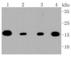
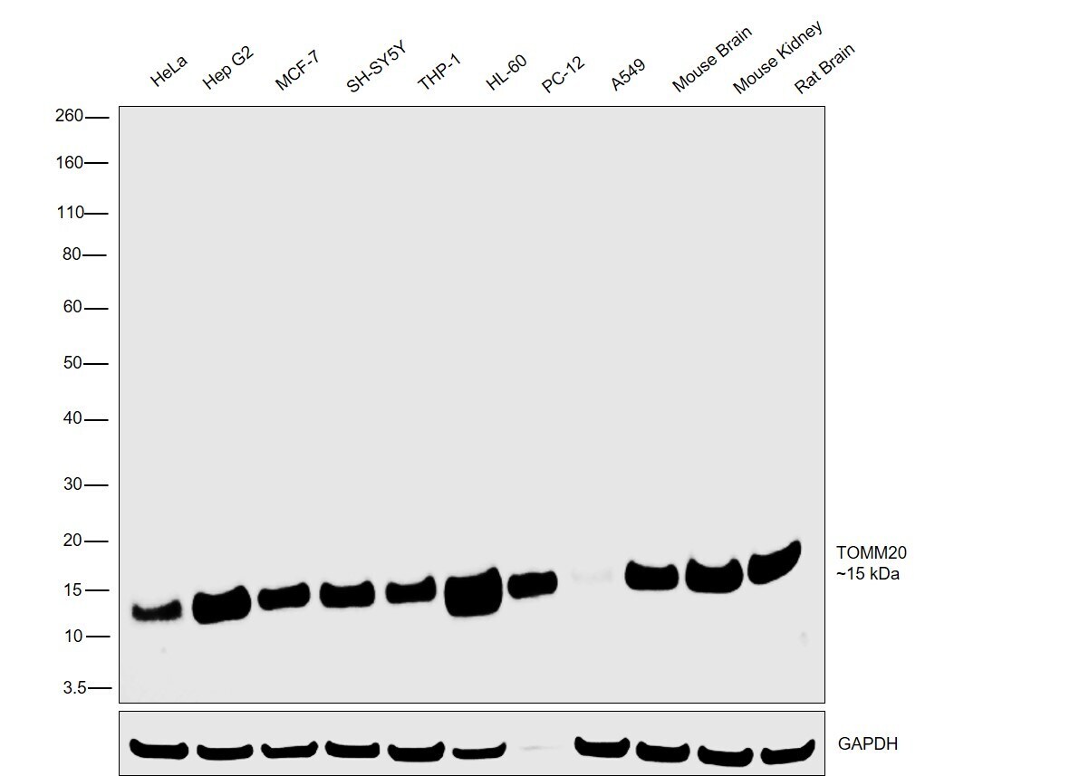
- Western blot analysis of TOMM20 in different lysates using a Monoclonal antibody (Product #MA5-32148) at a dilution of 1:1,000. Positive control: Lane 1: Hela, Lane 2: MCF-7, Lane 3: F9, Lane 4: PC12.

- Submitted by
- Invitrogen Antibodies (provider)
- Main image
- Experimental details
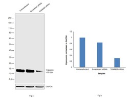
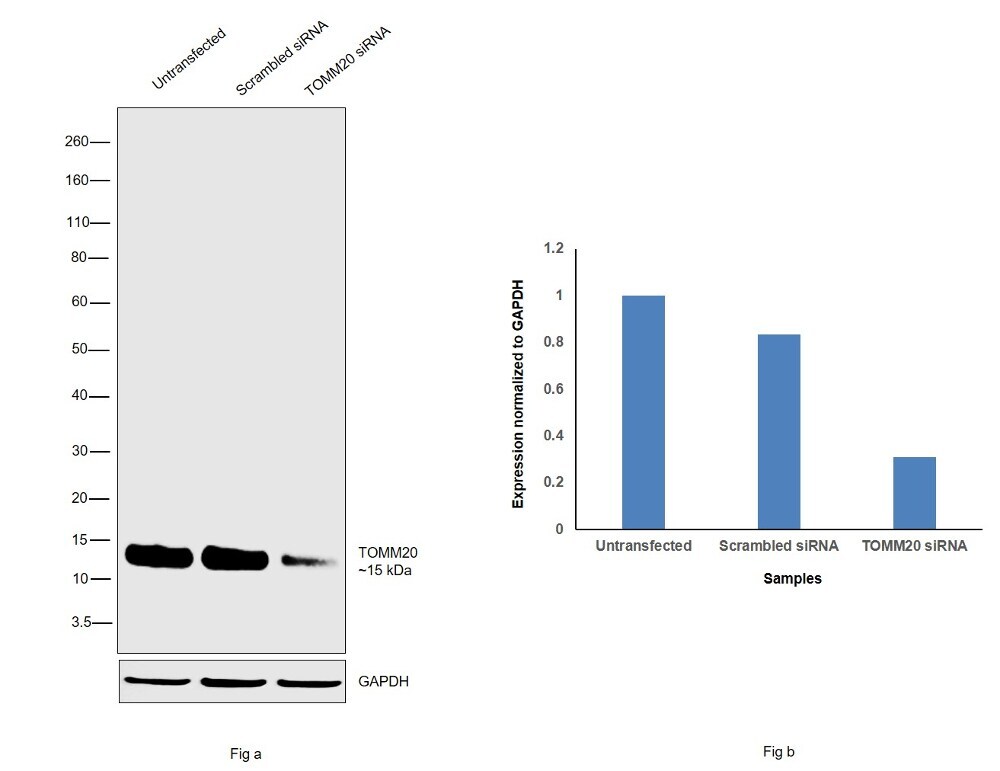
- Knockdown of TOMM20 was achieved by transfecting HeLa cells with TOMM20 specific siRNAs (Silencer® select Product # s18949, s18948). Western blot analysis (Fig. a) was performed using membrane extracts from the HeLa knockdown cells (lane 3), non-specific scrambled siRNA transfected cells (lane 2) and untransfected cells (lane 1). The blots were probed with TOMM20 Recombinant Rabbit Monoclonal Antibody (ST04-72) (Product # MA5-32148, 1:1,000 dilution) and Goat anti-Rabbit IgG (Heavy Chain), Superclonal™ Recombinant Secondary Antibody, HRP (Product # A27036, 0.25 µg/mL, 1:4,000 dilution). Densitometric analysis of this western blot is shown in histogram (Fig. b). Decrease in signal upon siRNA mediated knock down confirms that antibody is specific to TOMM20.
- Submitted by
- Invitrogen Antibodies (provider)
- Main image
- Experimental details
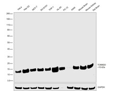
- Western blot was performed using Anti-TOMM20 Recombinant Rabbit Monoclonal Antibody (ST04-72) (Product # MA5-32148) and a 15kDa band corresponding to TOMM20 was observed across all the cell lines and tissue tested. Membrane extracts (30 µg lysate) of HeLa (Lane 1), Hep G2 (Lane 2), MCF-7 (Lane 3), SH-SY5Y (Lane 4), THP-1 (Lane 5), HL-60 (Lane 6), PC-12 (Lane 7), A549 (Lane 8), tissue extracts of Mouse Brain (Lane 9), Mouse Kidney (Lane 10) and Rat Brain (Lane 11) were electrophoresed using Novex® NuPAGE® 4-12% Bis-Tris Protein Gel (Product # NP0322BOX). Resolved proteins were then transferred onto a nitrocellulose membrane (Product # IB23001) by iBlot® 2 Dry Blotting System (Product # IB21001). The blot was probed with the primary antibody (1:1000 dilution) and detected by Goat anti-Rabbit IgG (Heavy Chain), Superclonal™ Recombinant Secondary Antibody, HRP (Product # A27036, 1:4000 dilution) using the iBright FL 1000 (Product # A32752). Chemiluminescent detection was performed using Novex® ECL Chemiluminescent Substrate Reagent Kit (Product # WP20005).
Supportive validation
- Submitted by
- Invitrogen Antibodies (provider)
- Main image
- Experimental details
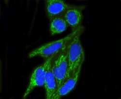
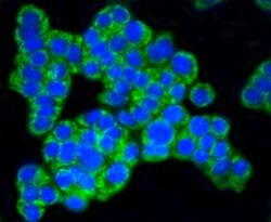
- Immunocytochemical analysis of TOMM20 in Hela cells using a TOMM20 Monoclonal antibody (Product # MA5-32148) as seen in green. The nuclear counter stain is DAPI (blue). Cells were fixed in paraformaldehyde, permeabilised with 0.25% Triton X100/PBS.

- Submitted by
- Invitrogen Antibodies (provider)
- Main image
- Experimental details
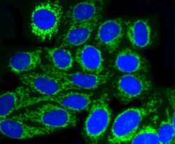
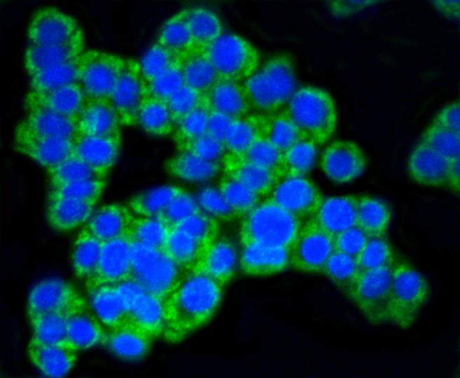
- Immunocytochemical analysis of TOMM20 in HepG2 cells using a TOMM20 Monoclonal antibody (Product # MA5-32148) as seen in green. The nuclear counter stain is DAPI (blue). Cells were fixed in paraformaldehyde, permeabilised with 0.25% Triton X100/PBS.

- Submitted by
- Invitrogen Antibodies (provider)
- Main image

- Experimental details
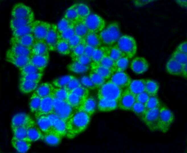
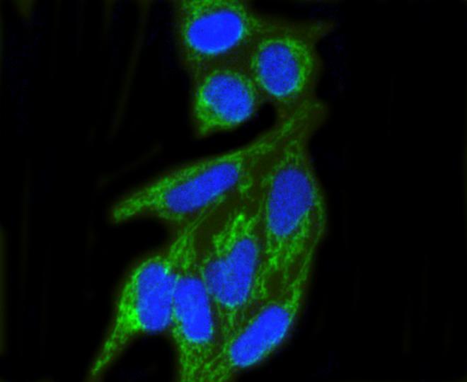
- Immunocytochemical analysis of TOMM20 in PC12 cells using a TOMM20 Monoclonal antibody (Product # MA5-32148) as seen in green. The nuclear counter stain is DAPI (blue). Cells were fixed in paraformaldehyde, permeabilised with 0.25% Triton X100/PBS.
- Submitted by
- Invitrogen Antibodies (provider)
- Main image

- Experimental details
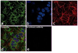
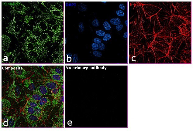
- Immunofluorescence analysis was performed using Anti-TOMM20 Rabbit Monoclonal Antibody (Product # MA5-32148) using 70% confluent log phase Hep G2 cells. The cells were fixed with 4% paraformaldehyde for 10 minutes, permeabilized with 0.1% Triton™ X-100 for 15 minutes, and blocked with 2% BSA for 1 hour at room temperature. The cells were labeled with TOMM20 Rabbit Monoclonal Antibody (Product # MA5-32148) at 1:100 dilution in 0.1% BSA and incubated at 4 degree Celsius overnight and then labeled with Goat anti-Rabbit IgG (H+L), Superclonal™ Recombinant Secondary Antibody, Alexa Fluor 488 (Product # A27034) at a dilution of 1:2000 for 45 minutes at room temperature (Panel a: green). Nuclei (Panel b: blue) were stained with ProLong™ Diamond Antifade Mountant with DAPI (Product # P36962). F-actin (Panel c: red) was stained with Rhodamine Phalloidin (Product # R415, 1:300). Panel d represents the merged image showing mitochondrial localization. Panel e represents the no primary control. The images were captured at 60X magnification.

- Submitted by
- Invitrogen Antibodies (provider)
- Main image
- Experimental details
- Immunocytochemical analysis of TOMM20 in PC12 cells using a TOMM20 Monoclonal antibody (Product # MA5-32148) as seen in green. The nuclear counter stain is DAPI (blue). Cells were fixed in paraformaldehyde, permeabilised with 0.25% Triton X100/PBS.
- Submitted by
- Invitrogen Antibodies (provider)
- Main image

- Experimental details
- Immunocytochemical analysis of TOMM20 in Hela cells using a TOMM20 Monoclonal antibody (Product # MA5-32148) as seen in green. The nuclear counter stain is DAPI (blue). Cells were fixed in paraformaldehyde, permeabilised with 0.25% Triton X100/PBS.
- Submitted by
- Invitrogen Antibodies (provider)
- Main image

- Experimental details
- Immunocytochemical analysis of TOMM20 in HepG2 cells using a TOMM20 Monoclonal antibody (Product # MA5-32148) as seen in green. The nuclear counter stain is DAPI (blue). Cells were fixed in paraformaldehyde, permeabilised with 0.25% Triton X100/PBS.

- Submitted by
- Invitrogen Antibodies (provider)
- Main image
- Experimental details
- Immunofluorescence analysis was performed using Anti-TOMM20 Rabbit Monoclonal Antibody (Product # MA5-32148) using 70% confluent log phase Hep G2 cells. The cells were fixed with 4% paraformaldehyde for 10 minutes, permeabilized with 0.1% Triton™ X-100 for 15 minutes, and blocked with 2% BSA for 1 hour at room temperature. The cells were labeled with TOMM20 Rabbit Monoclonal Antibody (Product # MA5-32148) at 1:100 dilution in 0.1% BSA and incubated at 4 degree Celsius overnight and then labeled with Goat anti-Rabbit IgG (Heavy Chain), Superclonal™ Recombinant Secondary Antibody, Alexa Fluor 488 (Product # A27034) at a dilution of 1:2000 for 45 minutes at room temperature (Panel a: green). Nuclei (Panel b: blue) were stained with ProLong™ Diamond Antifade Mountant with DAPI (Product # P36962). F-actin (Panel c: red) was stained with Rhodamine Phalloidin (Product # R415, 1:300). Panel d represents the merged image showing mitochondrial localization. Panel e represents the no primary control. The images were captured at 60X magnification.
Supportive validation
- Submitted by
- Invitrogen Antibodies (provider)
- Main image
- Experimental details
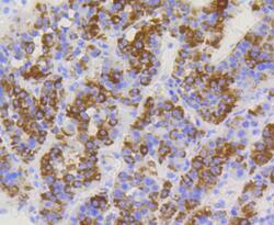
- Immunohistochemical analysis of TOMM20 of paraffin-embedded Human liver cancer tissue using a TOMM20 Monoclonal antibody (Product #MA5-32148). Counter stained with hematoxylin.

- Submitted by
- Invitrogen Antibodies (provider)
- Main image
- Experimental details
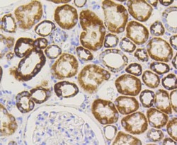
- Immunohistochemical analysis of TOMM20 of paraffin-embedded Human kidney tissue using a TOMM20 Monoclonal antibody (Product #MA5-32148). Counter stained with hematoxylin.

- Submitted by
- Invitrogen Antibodies (provider)
- Main image
- Experimental details
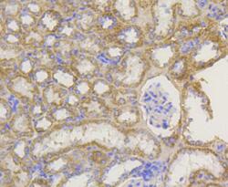
- Immunohistochemical analysis of TOMM20 of paraffin-embedded Mouse kidney tissue using a TOMM20 Monoclonal antibody (Product #MA5-32148). Counter stained with hematoxylin.

- Submitted by
- Invitrogen Antibodies (provider)
- Main image

- Experimental details
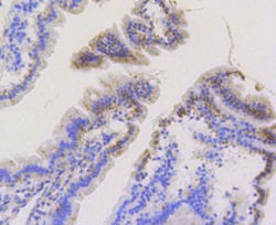
- Immunohistochemical analysis of TOMM20 of paraffin-embedded Mouse small intestine tissue using a TOMM20 Monoclonal antibody (Product #MA5-32148). Counter stained with hematoxylin. The nuclear counter stain is DAPI (blue).

- Submitted by
- Invitrogen Antibodies (provider)
- Main image
- Experimental details
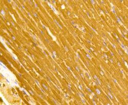
- Immunohistochemical analysis of TOMM20 of paraffin-embedded Mouse heart tissue using a TOMM20 Monoclonal antibody (Product #MA5-32148). Counter stained with hematoxylin.

- Submitted by
- Invitrogen Antibodies (provider)
- Main image
- Experimental details
- Immunohistochemical analysis of TOMM20 of paraffin-embedded Mouse small intestine tissue using a TOMM20 Monoclonal antibody (Product #MA5-32148). Counter stained with hematoxylin. The nuclear counter stain is DAPI (blue).

- Submitted by
- Invitrogen Antibodies (provider)
- Main image

- Experimental details
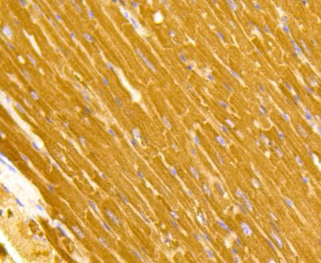
- Immunohistochemical analysis of TOMM20 of paraffin-embedded Mouse heart tissue using a TOMM20 Monoclonal antibody (Product #MA5-32148). Counter stained with hematoxylin.
Supportive validation
- Submitted by
- Invitrogen Antibodies (provider)
- Main image
- Experimental details
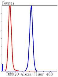
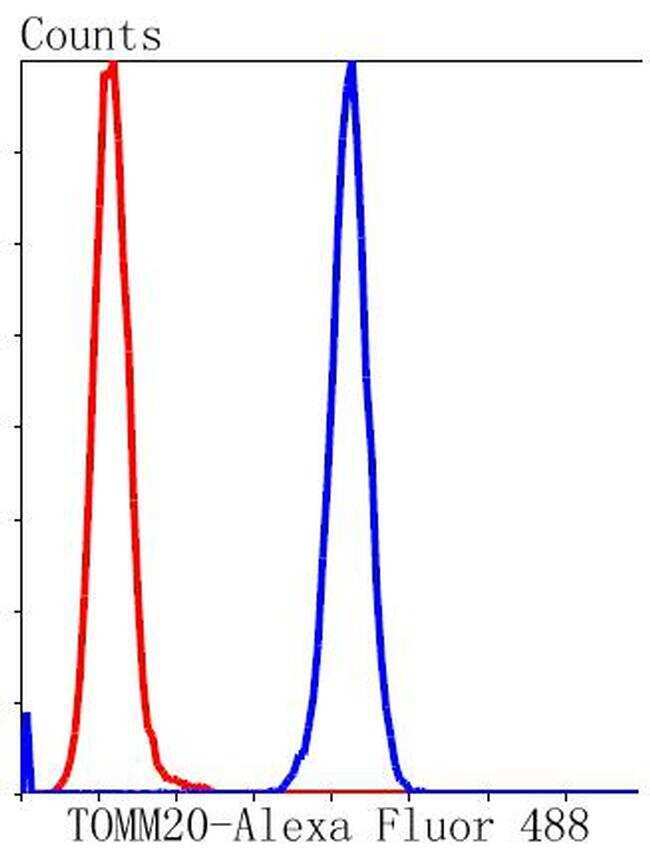
- Flow Cytometric analysis of TOMM20 in Hela cells using a TOMM20 Monoclonal Antibody (Product # MA5-32148) at a dilution of 1:50, as seen in blue compared with an unlabelled control (cells without incubation with primary antibody; red). Alexa Fluor 488-conjugated goat anti rabbit IgG was used as the secondary antibody.
- Submitted by
- Invitrogen Antibodies (provider)
- Main image
- Experimental details
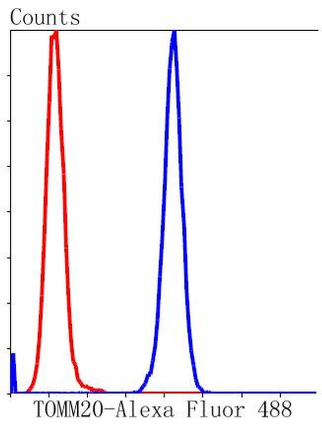
- Flow Cytometric analysis of TOMM20 in Hela cells using a TOMM20 Monoclonal Antibody (Product # MA5-32148) at a dilution of 1:50, as seen in blue compared with an unlabelled control (cells without incubation with primary antibody; red). Alexa Fluor 488-conjugated goat anti rabbit IgG was used as the secondary antibody.
Supportive validation
- Submitted by
- Invitrogen Antibodies (provider)
- Main image
- Experimental details
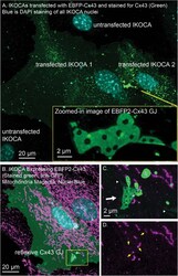
- FIGURE 6 IWCA and IKOCA cells are useful to study GJ morphology and interactions with other proteins with super-resolution microscopy. (A) IKOCA passage 10 cells transfected to re-express Cx43 with a fluorescent protein tag (green, immunolabeling with an antibody against Cx43) form GJs when two transfected cells are in contact. As indicated by blue DAPI staining, there are untransfected cells in contact with the two transfected IKOCA cells in the center of the field of the image in A that do not express Cx43 and do not form GJs (scale bar = 20 mum). Super-resolution imaging allows insights into structural features of the GJ plaque and endocytosed GJ plaque as shown in the zoomed inset (scale bar = 2 mum). (B) Re-expression of fluorescent protein tagged Cx43 in a subset of IKOCA cells with subsequent immunostaining for secondary cellular features present in all IKOCA cells (e.g., mitochondria) allows investigation of the effects of Cx43 expression in astrocyte-like cells within the same cell culture sample- facilitating side-by-side comparisons and visualization of interactions between Cx43 and other organelles with super-resolution microscopy (scale bar = 20 mum). (C) Zoomed in view of the location where a Cx43 GJ plaque structure (green, indicated by white arrow) appears to interact with small mitochondria (indicated by TOMM20 staining, magenta). This GJ plaque represents a reflexive GJ plaque that has formed where the plasma membrane of the same astrocyte overlaps to allow
