Explore
 Validate
 Learn
Learn Western blot
 ELISA
ELISA Immunoprecipitation
Antibody data
- Antibody Data
- Antigen structure
- References [0]
- Comments [0]
- Validations
- Western blot [3]
- Immunocytochemistry [2]
- Immunoprecipitation [2]
- Immunohistochemistry [11]
- Flow cytometry [2]
Submit
Validation data
Reference
Comment
Report error
- Product number
- RQ7209 - Provider product page

- Provider
- NSJ Bioreagents
- Product name
- TOMM20 Antibody
- Antibody type
- Polyclonal
- Description
- This highly specific TOMM20 antibody is suitable for use in Western blot/Immunohistochemistry/Immunofluorescence/Immunoprecipitation/Direct ELISA applications with human, mouse and rat samples.
- Reactivity
- Human, Mouse, Rat
- Host
- Rabbit
- Conjugate
- Unconjugated
- Vial size
- 100 ug
- Concentration
- 0.5mg/ml if reconstituted with 0.2ml sterile DI water
- Storage
- After reconstitution, the TOMM20 antibody can be stored for up to one month at 4oC. For long-term, aliquot and store at -20oC. Avoid repeated freezing and thawing.
No comments: Submit comment
Supportive validation
- Submitted by
- NSJ Bioreagents (provider)
- Main image
- Experimental details
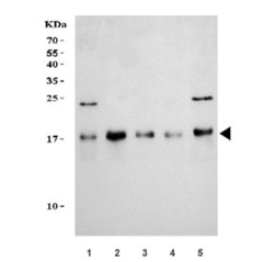
- Western blot testing of human 1) 293T, 2) HepG2, 3) HeLa, 4) SH-SY5Y and 5) HL60 cell lysate with TOMM20 antibody. Predicted molecular weight ~16 kDa.

- Submitted by
- NSJ Bioreagents (provider)
- Main image

- Experimental details
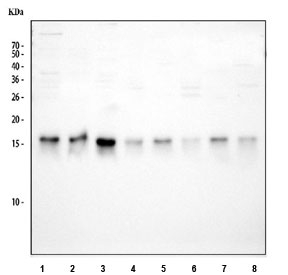
- Western blot testing of 1) human 293T, 2) human HeLa, 3) human HepG2, 4) human Jurkat, 5) rat brain, 6) rat liver, 7) mouse brain and 8) mouse liver tissue lysate with TOMM20 antibody. Predicted molecular weight ~16 kDa.
- Submitted by
- NSJ Bioreagents (provider)
- Main image

- Experimental details
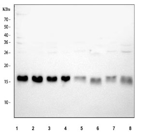
- Western blot testing of 1) human HepG2, 2) human 293T, 3) human K562, 4) human HeLa, 5) rat brain, 6) rat kidney, 7) mouse brain and 8) mouse kidney tissue lysate with TOMM20 antibody. Predicted molecular weight ~16 kDa.
Supportive validation
- Submitted by
- NSJ Bioreagents (provider)
- Main image

- Experimental details
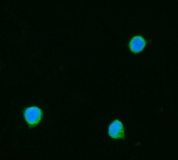
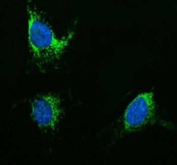
- Immunofluorescent staining of FFPE human SiHa cells with TOMM20 antibody (green) and DAPI nuclear stain (blue). HIER: steam section in pH6 citrate buffer for 20 min.
- Submitted by
- NSJ Bioreagents (provider)
- Main image

- Experimental details
- Immunofluorescent staining of FFPE human MG63 cells with TOMM20 antibody (green) and DAPI nuclear stain (blue). HIER: steam section in pH6 citrate buffer for 20 min.
Enhanced validation
- Submitted by
- NSJ Bioreagents (provider)
- Enhanced method
- Genetic validation
- Main image
- Experimental details
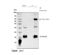
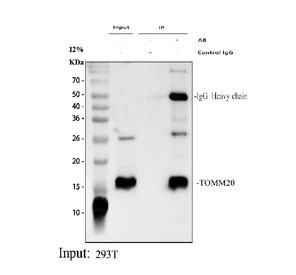
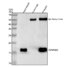
- Immunoprecipitation of TOMM20 protein from 500ug of human 293T whole cell lysate with 2ug of TOMM20 antibody.
- Submitted by
- NSJ Bioreagents (provider)
- Main image
- Experimental details
- Immunoprecipitation of TOMM20 protein from 500ug of human HepG2 whole cell lysate with 2ug of TOMM20 antibody.

Supportive validation
- Submitted by
- NSJ Bioreagents (provider)
- Main image
- Experimental details
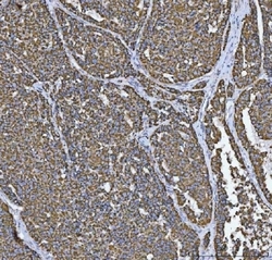
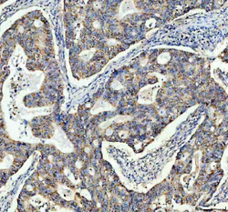
- IHC staining of FFPE human liver cancer tissue with TOMM20 antibody. HIER: boil tissue sections in pH8 EDTA for 20 min and allow to cool before testing.
- Submitted by
- NSJ Bioreagents (provider)
- Main image
- Experimental details
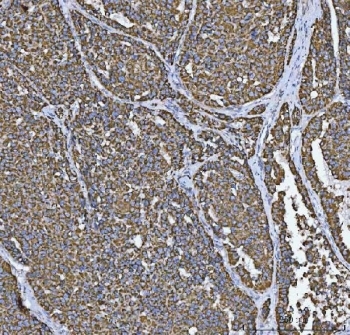
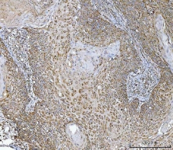
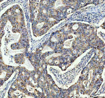
- IHC staining of FFPE human laryngeal squamous cell carcinoma tissue with TOMM20 antibody. HIER: boil tissue sections in pH8 EDTA for 20 min and allow to cool before testing.
- Submitted by
- NSJ Bioreagents (provider)
- Main image
- Experimental details
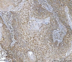
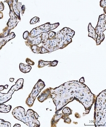
- IHC staining of FFPE human placental tissue with TOMM20 antibody. HIER: boil tissue sections in pH8 EDTA for 20 min and allow to cool before testing.

- Submitted by
- NSJ Bioreagents (provider)
- Main image
- Experimental details
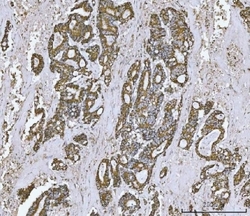
- IHC staining of FFPE human rectal cancer tissue with TOMM20 antibody. HIER: boil tissue sections in pH8 EDTA for 20 min and allow to cool before testing.

- Submitted by
- NSJ Bioreagents (provider)
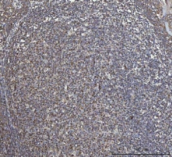
- Main image
- Experimental details
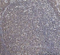
- IHC staining of FFPE human tonsil tissue with TOMM20 antibody. HIER: boil tissue sections in pH8 EDTA for 20 min and allow to cool before testing.
- Submitted by
- NSJ Bioreagents (provider)
- Main image
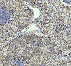
- Experimental details
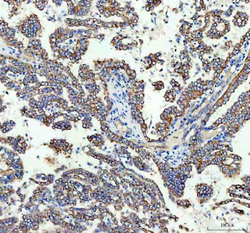
- IHC staining of FFPE human lung cancer tissue with TOMM20 antibody. HIER: boil tissue sections in pH8 EDTA for 20 min and allow to cool before testing.

- Submitted by
- NSJ Bioreagents (provider)
- Main image
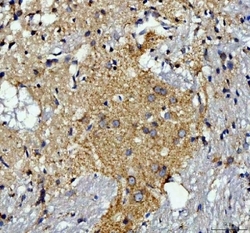
- Experimental details
- IHC staining of FFPE rat brain tissue with TOMM20 antibody. HIER: boil tissue sections in pH8 EDTA for 20 min and allow to cool before testing.

- Submitted by
- NSJ Bioreagents (provider)
- Main image
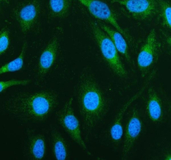
- Experimental details
- Immunofluorescent staining of FFPE human HeLa cells with TOMM20 antibody (green) and DAPI nuclear stain (blue). HIER: steam section in pH6 citrate buffer for 20 min.

- Submitted by
- NSJ Bioreagents (provider)
- Main image
- Experimental details
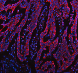
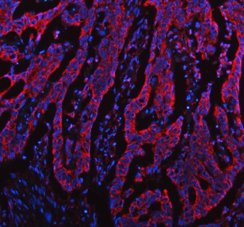
- Immunofluorescent staining of FFPE human colon cancer tissue with TOMM20 antibody (red) and DAPI nuclear stain (blue). HIER: steam section in pH8 EDTA buffer for 20 min.
- Submitted by
- NSJ Bioreagents (provider)
- Main image
- Experimental details
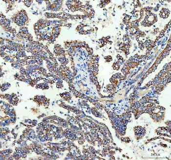
- IHC staining of FFPE human lung cancer tissue with TOMM20 antibody. HIER: boil tissue sections in pH8 EDTA for 20 min and allow to cool before testing.
- Submitted by
- NSJ Bioreagents (provider)
- Main image
- Experimental details
- IHC staining of FFPE human colon cancer tissue with TOMM20 antibody. HIER: boil tissue sections in pH8 EDTA for 20 min and allow to cool before testing.
Supportive validation
- Submitted by
- NSJ Bioreagents (provider)
- Main image
- Experimental details
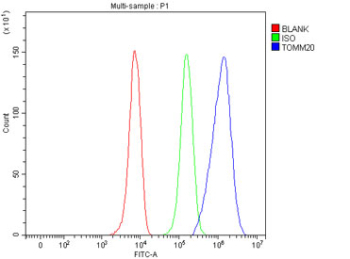
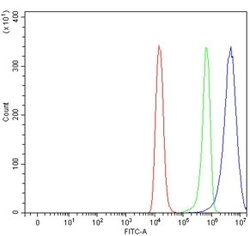
- Flow cytometry testing of human Raji cells with TOMM20 antibody at 1ug/million cells (blocked with goat sera); Red=cells alone, Green=isotype control, Blue= TOMM20 antibody.
- Submitted by
- NSJ Bioreagents (provider)
- Main image
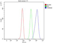
- Experimental details
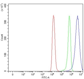
- Flow cytometry testing of fixed and permeabilized human HepG2 cells with TOMM20 antibody at 1ug/million cells (blocked with goat sera); Red=cells alone, Green=isotype control, Blue= TOMM20 antibody.