Explore
 Validate
 Learn
Learn Western blot
 Immunocytochemistry
Antibody data
- Antibody Data
- Antigen structure
- References [49]
- Comments [0]
- Validations
- Immunocytochemistry [1]
Submit
Validation data
Reference
Comment
Report error
- Product number
- HPA011562 - Provider product page

- Provider
- Atlas Antibodies
- Proper citation
- Atlas Antibodies Cat#HPA011562, RRID:AB_1080326
- Product name
- Anti-TOMM20
- Antibody type
- Polyclonal
- Description
- Polyclonal Antibody against Human TOMM20, Gene description: translocase of outer mitochondrial membrane 20 homolog (yeast), Alternative Gene Names: KIAA0016, MAS20, MOM19, TOM20, Validated applications: ICC, IHC, WB, Uniprot ID: Q15388, Storage: Store at +4°C for short term storage. Long time storage is recommended at -20°C.
- Reactivity
- Human
- Host
- Rabbit
- Conjugate
- Unconjugated
- Isotype
- IgG
- Vial size
- 100 µl
- Concentration
- 0.1 mg/ml
- Storage
- Store at +4°C for short term storage. Long time storage is recommended at -20°C.
- Handling
- The antibody solution should be gently mixed before use.
Submitted references The P2X7 Receptor is a Master Regulator of Microparticle and Mitochondria Exchange in Mouse Microglia
Acetalax (Oxyphenisatin Acetate, NSC 59687) and Bisacodyl Cause Oncosis in Triple-Negative Breast Cancer Cell Lines by Poisoning the Ion Exchange Membrane Protein TRPM4
TANGO2-related rhabdomyolysis symptoms are associated with abnormal autophagy functioning
MAPK1 Mediates MAM Disruption and Mitochondrial Dysfunction in Diabetic Kidney Disease via the PACS-2-Dependent Mechanism
Poly-GP accumulation due to C9orf72 loss of function induces motor neuron apoptosis through autophagy and mitophagy defects
Quantifying nanoscopic alterations associated with mitochondrial dysfunction using three-dimensional single-molecule localization microscopy
Neuronal extracellular vesicles influence the expression, degradation and oligomeric state of fructose 1,6-bisphosphatase 2 in astrocytes affecting their glycolytic capacity
Segregation of pathways leading to pexophagy
Mitochondrial rewiring drives metabolic adaptation to NAD(H) shortage in triple negative breast cancer cells
Activation of the cGAS-STING innate immune response in cells with deficient mitochondrial topoisomerase TOP1MT
A pesticide and iPSC dopaminergic neuron screen identifies and classifies Parkinson-relevant pesticides
Replication-associated formation and repair of human topoisomerase IIIα cleavage complexes.
Mitochondrial uncouplers induce proton leak by activating AAC and UCP1
Top3α is the replicative topoisomerase in mitochondrial DNA replication
GDAP1 loss of function inhibits the mitochondrial pyruvate dehydrogenase complex by altering the actin cytoskeleton
Functional characterization of two variants of mitochondrial topoisomerase TOP1MT that impact regulation of the mitochondrial genome
Intracellular IL-32 regulates mitochondrial metabolism, proliferation, and differentiation of malignant plasma cells
A novel mechanism for NLRP3 inflammasome activation
Extracellular ATP is increased by release of ATP-loaded microparticles triggered by nutrient deprivation
Stimulated emission depletion microscopy with a single depletion laser using five fluorochromes and fluorescence lifetime phasor separation
FBP2—A New Player in Regulation of Motility of Mitochondria and Stability of Microtubules in Cardiomyocytes
Self‐assembly of multi‐component mitochondrial nucleoids via phase separation
ATAD3A has a scaffolding role regulating mitochondria inner membrane structure and protein assembly
NIX initiates mitochondrial fragmentation via DRP1 to drive epidermal differentiation
The Tumor Necrosis Factor Alpha and Interleukin 6 Auto-paracrine Signaling Loop Controls Mycobacterium avium Infection via Induction of IRF1/IRG1 in Human Primary Macrophages
PERM1 interacts with the MICOS-MIB complex to connect the mitochondria and sarcolemma via ankyrin B
Characterization of a novel variant in the HR1 domain of MFN2 in a patient with ataxia, optic atrophy and sensorineural hearing loss
A Scaffold-Free 3-D Co-Culture Mimics the Major Features of the Reverse Warburg Effect In Vitro
Intact vitamin A transport is critical for cold-mediated adipose tissue browning and thermogenesis
USP30 sets a trigger threshold for PINK1–PARKIN amplification of mitochondrial ubiquitylation
A Splice Intervention Therapy for Autosomal Recessive Juvenile Parkinson’s Disease Arising from Parkin Mutations
Expression of Fbp2, a Newly Discovered Constituent of Memory Formation Mechanisms, Is Regulated by Astrocyte–Neuron Crosstalk
Functional Consequences of PDK4 Deficiency in Doberman Pinscher Fibroblasts
SS-31 Peptide Reverses the Mitochondrial Fragmentation Present in Fibroblasts From Patients With DCMA, a Mitochondrial Cardiomyopathy
Increased mtDNA Abundance and Improved Function in Human Barth Syndrome Patient Fibroblasts Following AAV-TAZ Gene Delivery
The zebrafish orthologue of the human hepatocerebral disease geneMPV17plays pleiotropic roles in mitochondria
Ciprofloxacin impairs mitochondrial DNA replication initiation through inhibition of Topoisomerase 2
PARL partitions the lipid transfer protein STARD7 between the cytosol and mitochondria
Pgam5 released from damaged mitochondria induces mitochondrial biogenesis via Wnt signaling
Dual role of USP 30 in controlling basal pexophagy and mitophagy
p62 is linked to mitophagy in oleic acid-induced adipogenesis in human adipose-derived stromal cells
The endogenous subcellular localisations of the long chain fatty acid-activating enzymes ACSL3 and ACSL4 in sarcoma and breast cancer cells.
Intracellular localization of polymyxins in human alveolar epithelial cells
PDK4 Deficiency Induces Intrinsic Apoptosis in Response to Starvation in Fibroblasts from Doberman Pinschers with Dilated Cardiomyopathy
New protein–protein interactions of mitochondrial connexin 43 in mouse heart
APOOL is a cardiolipin-binding constituent of the Mitofilin/MINOS protein complex determining cristae morphology in mammalian mitochondria.
APOOL Is a Cardiolipin-Binding Constituent of the Mitofilin/MINOS Protein Complex Determining Cristae Morphology in Mammalian Mitochondria
Systematic validation of antibody binding and protein subcellular localization using siRNA and confocal microscopy
Falzoni S, Vultaggio-Poma V, Chiozzi P, Tarantini M, Adinolfi E, Boldrini P, Giuliani A, Morciano G, Tang Y, Gorecki D, Di Virgilio F
Function 2024;5(4)
Function 2024;5(4)
Acetalax (Oxyphenisatin Acetate, NSC 59687) and Bisacodyl Cause Oncosis in Triple-Negative Breast Cancer Cell Lines by Poisoning the Ion Exchange Membrane Protein TRPM4
Mizunuma M, Redon C, Saha L, Tran A, Dhall A, Sebastian R, Taniyama D, Kruhlak M, Reinhold W, Takebe N, Pommier Y
Cancer Research Communications 2024;4(8):2101-2111
Cancer Research Communications 2024;4(8):2101-2111
TANGO2-related rhabdomyolysis symptoms are associated with abnormal autophagy functioning
de Calbiac H, Montealegre S, Straube M, Renault S, Debruge H, Chentout L, Ciura S, Imbard A, Le Guillou E, Marian A, Goudin N, Caccavelli L, Fabrega S, Hubas A, van Endert P, Dupont N, Diana J, Kabashi E, de Lonlay P
Autophagy Reports 2024;3(1)
Autophagy Reports 2024;3(1)
MAPK1 Mediates MAM Disruption and Mitochondrial Dysfunction in Diabetic Kidney Disease via the PACS-2-Dependent Mechanism
Liu S, Han S, Wang C, Chen H, Xu Q, Feng S, Wang Y, Yao J, Zhou Q, Tang X, Lin L, Hu L, Davidson A, Yang B, Ye C, Yang F, Mao J, Tong C, Chen J, Jiang H
International Journal of Biological Sciences 2024;20(2):569-584
International Journal of Biological Sciences 2024;20(2):569-584
Poly-GP accumulation due to C9orf72 loss of function induces motor neuron apoptosis through autophagy and mitophagy defects
de Calbiac H, Renault S, Haouy G, Jung V, Roger K, Zhou Q, Campanari M, Chentout L, Demy D, Marian A, Goudin N, Edbauer D, Guerrera C, Ciura S, Kabashi E
Autophagy 2024;20(10):2164-2185
Autophagy 2024;20(10):2164-2185
Quantifying nanoscopic alterations associated with mitochondrial dysfunction using three-dimensional single-molecule localization microscopy
Brenner B, Xu F, Zhang Y, Kweon J, Fang R, Sheibani N, Zhang S, Sun C, Zhang H
Biomedical Optics Express 2024;15(3):1571
Biomedical Optics Express 2024;15(3):1571
Neuronal extracellular vesicles influence the expression, degradation and oligomeric state of fructose 1,6-bisphosphatase 2 in astrocytes affecting their glycolytic capacity
Hajka D, Budziak B, Rakus D, Gizak A
Scientific Reports 2024;14(1)
Scientific Reports 2024;14(1)
Yeo W, Brenner B, Lee Y, Kweon J, Sun C, Zhang H
2024
2024
Segregation of pathways leading to pexophagy
Barone F, Urbé S, Clague M
Life Science Alliance 2023;6(5):e202201825
Life Science Alliance 2023;6(5):e202201825
Mitochondrial rewiring drives metabolic adaptation to NAD(H) shortage in triple negative breast cancer cells
Carreira A, Ravera S, Zucal C, Thongon N, Irene C, Astigiano C, Bertola N, Buongiorno A, Roccuzzo M, Bisio A, Pardini B, Nencioni A, Bruzzone S, Provenzani A
Neoplasia 2023;41
Neoplasia 2023;41
Activation of the cGAS-STING innate immune response in cells with deficient mitochondrial topoisomerase TOP1MT
Al Khatib I, Deng J, Lei Y, Torres-Odio S, Rojas G, Newman L, Chung B, Symes A, Zhang H, Huang S, Pommier Y, Khan A, Shadel G, West A, Gibson W, Shutt T
Human Molecular Genetics 2023;32(15):2422-2440
Human Molecular Genetics 2023;32(15):2422-2440
A pesticide and iPSC dopaminergic neuron screen identifies and classifies Parkinson-relevant pesticides
Paul K, Krolewski R, Lucumi Moreno E, Blank J, Holton K, Ahfeldt T, Furlong M, Yu Y, Cockburn M, Thompson L, Kreymerman A, Ricci-Blair E, Li Y, Patel H, Lee R, Bronstein J, Rubin L, Khurana V, Ritz B
Nature Communications 2023;14(1)
Nature Communications 2023;14(1)
Replication-associated formation and repair of human topoisomerase IIIα cleavage complexes.
Saha LK, Saha S, Yang X, Huang SN, Sun Y, Jo U, Pommier Y
Nature communications 2023 Apr 6;14(1):1925
Nature communications 2023 Apr 6;14(1):1925
Mitochondrial uncouplers induce proton leak by activating AAC and UCP1
Bertholet A, Natale A, Bisignano P, Suzuki J, Fedorenko A, Hamilton J, Brustovetsky T, Kazak L, Garrity R, Chouchani E, Brustovetsky N, Grabe M, Kirichok Y
Nature 2022;606(7912):180-187
Nature 2022;606(7912):180-187
Top3α is the replicative topoisomerase in mitochondrial DNA replication
Hangas A, Kekäläinen N, Potter A, Michell C, Aho K, Rutanen C, Spelbrink J, Pohjoismäki J, Goffart S
Nucleic Acids Research 2022;50(15):8733-8748
Nucleic Acids Research 2022;50(15):8733-8748
GDAP1 loss of function inhibits the mitochondrial pyruvate dehydrogenase complex by altering the actin cytoskeleton
Wolf C, Pouya A, Bitar S, Pfeiffer A, Bueno D, Rojas-Charry L, Arndt S, Gomez-Zepeda D, Tenzer S, Bello F, Vianello C, Ritz S, Schwirz J, Dobrindt K, Peitz M, Hanschmann E, Mencke P, Boussaad I, Silies M, Brüstle O, Giacomello M, Krüger R, Methner A
Communications Biology 2022;5(1)
Communications Biology 2022;5(1)
Functional characterization of two variants of mitochondrial topoisomerase TOP1MT that impact regulation of the mitochondrial genome
Al Khatib I, Deng J, Symes A, Kerr M, Zhang H, Huang S, Pommier Y, Khan A, Shutt T
Journal of Biological Chemistry 2022;298(10):102420
Journal of Biological Chemistry 2022;298(10):102420
Intracellular IL-32 regulates mitochondrial metabolism, proliferation, and differentiation of malignant plasma cells
Aass K, Mjelle R, Kastnes M, Tryggestad S, van den Brink L, Aass Roseth I, Westhrin M, Zahoor M, Moen S, Vikene Nedal T, Buene G, Misund K, Sponaas A, Ma Q, Sundan A, Groen R, Slørdahl T, Waage A, Standal T
iScience 2022;25(1):103605
iScience 2022;25(1):103605
A novel mechanism for NLRP3 inflammasome activation
Zhang T, Zhao J, Liu T, Cheng W, Wang Y, Ding S, Wang R
Metabolism Open 2022;13
Metabolism Open 2022;13
Extracellular ATP is increased by release of ATP-loaded microparticles triggered by nutrient deprivation
Vultaggio-Poma V, Falzoni S, Chiozzi P, Sarti A, Adinolfi E, Giuliani A, Sánchez-Melgar A, Boldrini P, Zanoni M, Tesei A, Pinton P, Di Virgilio F
Theranostics 2022;12(2):859-874
Theranostics 2022;12(2):859-874
Stimulated emission depletion microscopy with a single depletion laser using five fluorochromes and fluorescence lifetime phasor separation
Gonzalez Pisfil M, Nadelson I, Bergner B, Rottmeier S, Thomae A, Dietzel S
Scientific Reports 2022;12(1)
Scientific Reports 2022;12(1)
FBP2—A New Player in Regulation of Motility of Mitochondria and Stability of Microtubules in Cardiomyocytes
Pietras Ł, Stefanik E, Rakus D, Gizak A
Cells 2022;11(10):1710
Cells 2022;11(10):1710
Self‐assembly of multi‐component mitochondrial nucleoids via phase separation
Feric M, Demarest T, Tian J, Croteau D, Bohr V, Misteli T
The EMBO Journal 2021;40(6)
The EMBO Journal 2021;40(6)
ATAD3A has a scaffolding role regulating mitochondria inner membrane structure and protein assembly
Arguello T, Peralta S, Antonicka H, Gaidosh G, Diaz F, Tu Y, Garcia S, Shiekhattar R, Barrientos A, Moraes C
Cell Reports 2021;37(12):110139
Cell Reports 2021;37(12):110139
NIX initiates mitochondrial fragmentation via DRP1 to drive epidermal differentiation
Simpson C, Tokito M, Uppala R, Sarkar M, Gudjonsson J, Holzbaur E
Cell Reports 2021;34(5):108689
Cell Reports 2021;34(5):108689
The Tumor Necrosis Factor Alpha and Interleukin 6 Auto-paracrine Signaling Loop Controls Mycobacterium avium Infection via Induction of IRF1/IRG1 in Human Primary Macrophages
Gidon A, Louet C, Røst L, Bruheim P, Flo T, Stallings C
mBio 2021;12(5)
mBio 2021;12(5)
PERM1 interacts with the MICOS-MIB complex to connect the mitochondria and sarcolemma via ankyrin B
Bock T, Türk C, Aravamudhan S, Keufgens L, Bloch W, Rozsivalova D, Romanello V, Nogara L, Blaauw B, Trifunovic A, Braun T, Krüger M
Nature Communications 2021;12(1)
Nature Communications 2021;12(1)
Characterization of a novel variant in the HR1 domain of MFN2 in a patient with ataxia, optic atrophy and sensorineural hearing loss
Sharma G, Sabouny R, Joel M, Martens K, Martino D, de Koning A, Pfeffer G, Shutt T
F1000Research 2021;10
F1000Research 2021;10
A Scaffold-Free 3-D Co-Culture Mimics the Major Features of the Reverse Warburg Effect In Vitro
Keller F, Bruch R, Schneider R, Meier-Hubberten J, Hafner M, Rudolf R
Cells 2020;9(8):1900
Cells 2020;9(8):1900
Intact vitamin A transport is critical for cold-mediated adipose tissue browning and thermogenesis
Fenzl A, Kulterer O, Spirk K, Mitulović G, Marculescu R, Bilban M, Baumgartner-Parzer S, Kautzky-Willer A, Kenner L, Plutzky J, Quadro L, Kiefer F
Molecular Metabolism 2020;42
Molecular Metabolism 2020;42
USP30 sets a trigger threshold for PINK1–PARKIN amplification of mitochondrial ubiquitylation
Rusilowicz-Jones E, Jardine J, Kallinos A, Pinto-Fernandez A, Guenther F, Giurrandino M, Barone F, McCarron K, Burke C, Murad A, Martinez A, Marcassa E, Gersch M, Buckmelter A, Kayser-Bricker K, Lamoliatte F, Gajbhiye A, Davis S, Scott H, Murphy E, England K, Mortiboys H, Komander D, Trost M, Kessler B, Ioannidis S, Ahlijanian M, Urbé S, Clague M
Life Science Alliance 2020;3(8):e202000768
Life Science Alliance 2020;3(8):e202000768
A Splice Intervention Therapy for Autosomal Recessive Juvenile Parkinson’s Disease Arising from Parkin Mutations
Li D, Aung-Htut M, Ham K, Fletcher S, Wilton S
International Journal of Molecular Sciences 2020;21(19):7282
International Journal of Molecular Sciences 2020;21(19):7282
Expression of Fbp2, a Newly Discovered Constituent of Memory Formation Mechanisms, Is Regulated by Astrocyte–Neuron Crosstalk
Hajka D, Duda P, Wójcicka O, Drulis-Fajdasz D, Rakus D, Gizak A
International Journal of Molecular Sciences 2020;21(18):6903
International Journal of Molecular Sciences 2020;21(18):6903
Functional Consequences of PDK4 Deficiency in Doberman Pinscher Fibroblasts
Bolfer L, Estrada A, Larkin C, Conlon T, Lourenco F, Taggart K, Suzuki-Hatano S, Pacak C
Scientific Reports 2020;10(1)
Scientific Reports 2020;10(1)
SS-31 Peptide Reverses the Mitochondrial Fragmentation Present in Fibroblasts From Patients With DCMA, a Mitochondrial Cardiomyopathy
Machiraju P, Wang X, Sabouny R, Huang J, Zhao T, Iqbal F, King M, Prasher D, Lodha A, Jimenez-Tellez N, Ravandi A, Argiropoulos B, Sinasac D, Khan A, Shutt T, Greenway S
Frontiers in Cardiovascular Medicine 2019;6
Frontiers in Cardiovascular Medicine 2019;6
Increased mtDNA Abundance and Improved Function in Human Barth Syndrome Patient Fibroblasts Following AAV-TAZ Gene Delivery
Suzuki-Hatano S, Sriramvenugopal M, Ramanathan M, Soustek M, Byrne B, Cade W, Kang P, Pacak C
International Journal of Molecular Sciences 2019;20(14):3416
International Journal of Molecular Sciences 2019;20(14):3416
The zebrafish orthologue of the human hepatocerebral disease geneMPV17plays pleiotropic roles in mitochondria
Martorano L, Peron M, Laquatra C, Lidron E, Facchinello N, Meneghetti G, Tiso N, Rasola A, Ghezzi D, Argenton F
Disease Models & Mechanisms 2019
Disease Models & Mechanisms 2019
Ciprofloxacin impairs mitochondrial DNA replication initiation through inhibition of Topoisomerase 2
Hangas A, Aasumets K, Kekäläinen N, Paloheinä M, Pohjoismäki J, Gerhold J, Goffart S
Nucleic Acids Research 2018;46(18):9625-9636
Nucleic Acids Research 2018;46(18):9625-9636
PARL partitions the lipid transfer protein STARD7 between the cytosol and mitochondria
Saita S, Tatsuta T, Lampe P, König T, Ohba Y, Langer T
The EMBO Journal 2018;37(4)
The EMBO Journal 2018;37(4)
Pgam5 released from damaged mitochondria induces mitochondrial biogenesis via Wnt signaling
Bernkopf D, Jalal K, Brückner M, Knaup K, Gentzel M, Schambony A, Behrens J
Journal of Cell Biology 2018;217(4):1383-1394
Journal of Cell Biology 2018;217(4):1383-1394
Dual role of USP 30 in controlling basal pexophagy and mitophagy
Marcassa E, Kallinos A, Jardine J, Rusilowicz‐Jones E, Martinez A, Kuehl S, Islinger M, Clague M, Urbé S
EMBO reports 2018;19(7)
EMBO reports 2018;19(7)
p62 is linked to mitophagy in oleic acid-induced adipogenesis in human adipose-derived stromal cells
Zeng R, Fang Y, Zhang Y, Bai S
Lipids in Health and Disease 2018;17(1)
Lipids in Health and Disease 2018;17(1)
The endogenous subcellular localisations of the long chain fatty acid-activating enzymes ACSL3 and ACSL4 in sarcoma and breast cancer cells.
Radif Y, Ndiaye H, Kalantzi V, Jacobs R, Hall A, Minogue S, Waugh MG
Molecular and cellular biochemistry 2018 Nov;448(1-2):275-286
Molecular and cellular biochemistry 2018 Nov;448(1-2):275-286
Intracellular localization of polymyxins in human alveolar epithelial cells
Ahmed M, Velkov T, Zhou Q, Fulcher A, Callaghan J, Zhou F, Chan K, Azad M, Li J
Journal of Antimicrobial Chemotherapy 2018
Journal of Antimicrobial Chemotherapy 2018
PDK4 Deficiency Induces Intrinsic Apoptosis in Response to Starvation in Fibroblasts from Doberman Pinschers with Dilated Cardiomyopathy
Taggart K, Estrada A, Thompson P, Lourenco F, Kirmani S, Suzuki-Hatano S, Pacak C
BioResearch Open Access 2017;6(1):182-191
BioResearch Open Access 2017;6(1):182-191
New protein–protein interactions of mitochondrial connexin 43 in mouse heart
Denuc A, Núñez E, Calvo E, Loureiro M, Miro‐Casas E, Guarás A, Vázquez J, Garcia‐Dorado D
Journal of Cellular and Molecular Medicine 2016;20(5):794-803
Journal of Cellular and Molecular Medicine 2016;20(5):794-803
APOOL is a cardiolipin-binding constituent of the Mitofilin/MINOS protein complex determining cristae morphology in mammalian mitochondria.
Weber TA, Koob S, Heide H, Wittig I, Head B, van der Bliek A, Brandt U, Mittelbronn M, Reichert AS
PloS one 2013;8(5):e63683
PloS one 2013;8(5):e63683
APOOL Is a Cardiolipin-Binding Constituent of the Mitofilin/MINOS Protein Complex Determining Cristae Morphology in Mammalian Mitochondria
Weber T, Koob S, Heide H, Wittig I, Head B, van der Bliek A, Brandt U, Mittelbronn M, Reichert A, Cobine P
PLoS ONE 2013 May;8(5)
PLoS ONE 2013 May;8(5)
Systematic validation of antibody binding and protein subcellular localization using siRNA and confocal microscopy
Stadler C, Hjelmare M, Neumann B, Jonasson K, Pepperkok R, Uhlén M, Lundberg E
Journal of Proteomics 2012;75(7):2236-2251
Journal of Proteomics 2012;75(7):2236-2251
No comments: Submit comment
Supportive validation
- Submitted by
- Atlas Antibodies (provider)
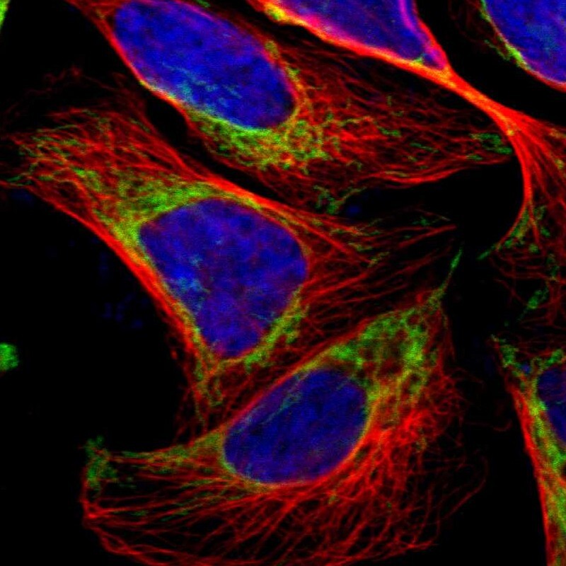
- Main image

- Experimental details
- Immunofluorescent staining of human cell line U-2 OS shows localization to mitochondria.
- Sample type
- Human