 Explore
 Validate
 Learn
Learn Western blot
Antibody data
- Antibody Data
- Antigen structure
- References [0]
- Comments [0]
- Validations
- Western blot [1]
- Immunocytochemistry [2]
- Immunohistochemistry [2]
- Flow cytometry [2]
Submit
Validation data
Reference
Comment
Report error
- Product number
- PA5-26030 - Provider product page

- Provider
- Invitrogen Antibodies
- Product name
- JIP1 Polyclonal Antibody
- Antibody type
- Polyclonal
- Antigen
- Synthetic peptide
- Reactivity
- Human
- Host
- Rabbit
- Isotype
- IgG
- Vial size
- 400 μL
- Concentration
- 0.45 mg/mL
- Storage
- Store at 4°C short term. For long term storage, store at -20°C, avoiding freeze/thaw cycles.
No comments: Submit comment
Supportive validation
- Submitted by
- Invitrogen Antibodies (provider)
- Main image
- Experimental details
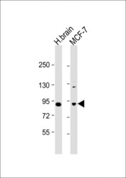
- Western blot analysis of JIP1 in various lysates. Samples were incubated with JIP1 polyclonal antibody (Product # PA5-26030) using a dilution of 1:1,000 followed by Goat Anti-Rabbit IgG, (H+L), Peroxidase conjugated at a dilution of 1:10,000. Lysates/proteins: 20 µg per lane. Lane 1: human brain lysate; Lane 2: MCF-7 whole cell lysate. Predicted band size: 78 kDa. Blocking/Dilution buffer: 5% NFDM/TBST.

Supportive validation
- Submitted by
- Invitrogen Antibodies (provider)
- Main image
- Experimental details
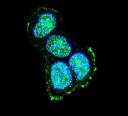
- Immunofluorescent analysis of MCF-7 cells using a JIP1 polyclonal antibody (Product # PA5-26030) at a dilution of 1:10-50, followed by a fluor-conjugated goat anti-rabbit secondary antibody (green). Nuclei were stained with DAPI (blue).

- Submitted by
- Invitrogen Antibodies (provider)
- Main image

- Experimental details
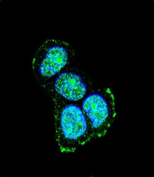
- Immunocytochemistry analysis of JIP1 in MCF-7 cells. Samples were incubated in JIP1 polyclonal antibody (Product # PA5-26030) followed by Alexa Fluor® 488-conjugated goat anti-rabbit lgG (green). DAPI was used to stain the cell nuclear (blue).
Supportive validation
- Submitted by
- Invitrogen Antibodies (provider)
- Main image
- Experimental details
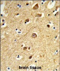
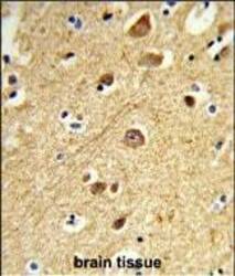
- Immunohistochemistry analysis of JIP1 in formalin-fixed and paraffin-embedded human brain tissue. Samples were incubated with JIP1 polyclonal antibody (Product # PA5-26030) which was peroxidase-conjugated to the secondary antibody, followed by DAB staining. This data demonstrates the use of this antibody for immunohistochemistry; clinical relevance has not been evaluated.

- Submitted by
- Invitrogen Antibodies (provider)
- Main image
- Experimental details
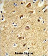
- Immunohistochemistry analysis of JIP1 in formalin-fixed and paraffin-embedded human brain tissue. Samples were incubated with JIP1 polyclonal antibody (Product # PA5-26030) which was peroxidase-conjugated to the secondary antibody, followed by DAB staining. This data demonstrates the use of this antibody for immunohistochemistry; clinical relevance has not been evaluated.
Supportive validation
- Submitted by
- Invitrogen Antibodies (provider)
- Main image
- Experimental details
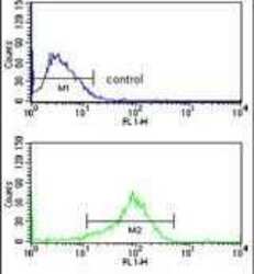
- Flow cytometry analysis of MCF-7 cells using a JIP1 polyclonal antibody (Product # PA5-26030) (bottom) compared to a negative control cell (top) at a dilution of 1:10-50, followed by a FITC-conjugated goat anti-rabbit antibody
- Submitted by
- Invitrogen Antibodies (provider)
- Main image
- Experimental details
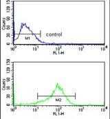
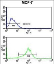
- Flow cytometry of JIP1 in MCF-7 cells (bottom histogram). Samples were incubated with JIP1 polyclonal antibody (Product # PA5-26030) followed by FITC-conjugated goat-anti-rabbit secondary antibody. Negative control cell (top histogram).
