Explore
 Validate
 Learn
Learn Western blot
Antibody data
- Antibody Data
- Antigen structure
- References [2]
- Comments [0]
- Validations
- Western blot [1]
- Immunohistochemistry [1]
- Other assay [1]
Submit
Validation data
Reference
Comment
Report error
- Product number
- PA5-42593 - Provider product page

- Provider
- Invitrogen Antibodies
- Product name
- GP2 Polyclonal Antibody
- Antibody type
- Polyclonal
- Antigen
- Synthetic peptide
- Description
- Peptide sequence: SLQAALQPIV SSLNVSVDGN GEFIVRMALF QDQNYTNPYE GDAVELSVES Sequence homology: Cow: 79%; Horse: 92%; Human: 100%; Mouse: 92%; Pig: 79%; Rat: 92%
- Reactivity
- Human
- Host
- Rabbit
- Isotype
- IgG
- Vial size
- 100 μL
- Concentration
- 0.5 mg/mL
- Storage
- -20°C, Avoid Freeze/Thaw Cycles
Submitted references Treatment with Commonly Used Antiretroviral Drugs Induces a Type I/III Interferon Signature in the Gut in the Absence of HIV Infection.
Ex Vivo and in Vivo Study of Sucrosomial(®) Iron Intestinal Absorption and Bioavailability.
Hughes SM, Levy CN, Calienes FL, Stekler JD, Pandey U, Vojtech L, Berard AR, Birse K, Noël-Romas L, Richardson B, Golden JB, Cartwright M, Collier AC, Stevens CE, Curlin ME, Holtz TH, Mugo N, Irungu E, Katabira E, Muwonge T, Lama JR, Baeten JM, Burgener A, Lingappa JR, McElrath MJ, Mackelprang R, McGowan I, Cranston RD, Cameron MJ, Hladik F
Cell reports. Medicine 2020 Sep 22;1(6):100096
Cell reports. Medicine 2020 Sep 22;1(6):100096
Ex Vivo and in Vivo Study of Sucrosomial(®) Iron Intestinal Absorption and Bioavailability.
Fabiano A, Brilli E, Mattii L, Testai L, Moscato S, Citi V, Tarantino G, Zambito Y
International journal of molecular sciences 2018 Sep 12;19(9)
International journal of molecular sciences 2018 Sep 12;19(9)
No comments: Submit comment
Supportive validation
- Submitted by
- Invitrogen Antibodies (provider)
- Main image

- Experimental details
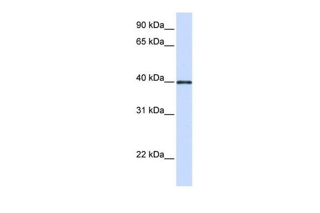
- Western blot analysis of human heart cells using an anti-GP2 polyclonal antibody (Product # PA5-42593).
Supportive validation
- Submitted by
- Invitrogen Antibodies (provider)
- Main image

- Experimental details
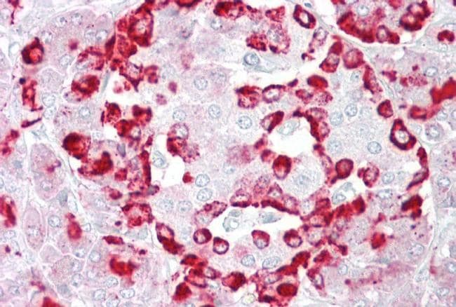
- Immunohistochemistry (paraffin-embedded) analysis of human pancreas tissue using an anti-GP2 polyclonal antibody (Product # PA5-42593).
Supportive validation
- Submitted by
- Invitrogen Antibodies (provider)
- Main image
- Experimental details
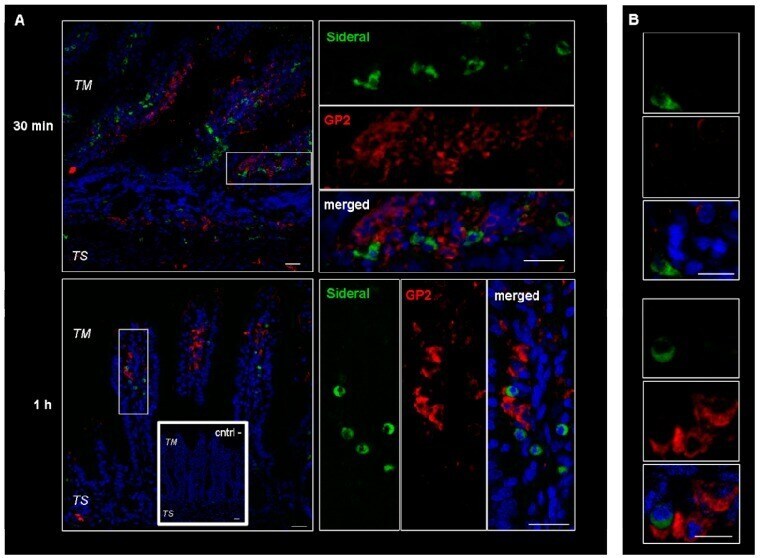
- Figure 4 Confocal laser scanning microscopy: representative images of rat gut sections after 0.5 and 1 h incubation in Ussing chamber. Negative controls obtained omitting primary antibodies are shown in double line squares. Individual fluorescent channels: green (SRM), red (GP2 + cells) and blue (nuclei). ( A ) bidimensional image of the maximum intensity projection. Scale bar 25 um. ( B ) Images acquired in one Z-plane (38th of 59, 22nd of 64, incubated 0.5 h and 1 h, respectively) at a higher magnification. Scale bar 10 um.