 Explore
 Validate
 Learn
Learn Western blot
Antibody data
- Antibody Data
- Antigen structure
- References [1]
- Comments [0]
- Validations
- Western blot [1]
- Flow cytometry [1]
Submit
Validation data
Reference
Comment
Report error
- Product number
- MAB63341 - Provider product page

- Provider
- Novus Biologicals
- Product name
- Mouse Monoclonal LAT Antibody
- Antibody type
- Monoclonal
- Description
- Protein A or G purified from hybridoma culture supernatant. Detects human LAT in direct ELISAs.
- Reactivity
- Human
- Host
- Mouse
- Isotype
- IgG
- Vial size
- 100 ug
- Concentration
- LYOPH
- Storage
- Use a manual defrost freezer and avoid repeated freeze-thaw cycles. 12 months from date of receipt, -20 to -70 degreesC as supplied. 1 month, 2 to 8 degreesC under sterile conditions after reconstitution. 6 months, -20 to -70 degreesC under sterile conditions after reconstitution.
Submitted references Rab6-dependent retrograde traffic of LAT controls immune synapse formation and T cell activation.
Carpier JM, Zucchetti AE, Bataille L, Dogniaux S, Shafaq-Zadah M, Bardin S, Lucchino M, Maurin M, Joannas LD, Magalhaes JG, Johannes L, Galli T, Goud B, Hivroz C
The Journal of experimental medicine 2018 Apr 2;215(4):1245-1265
The Journal of experimental medicine 2018 Apr 2;215(4):1245-1265
No comments: Submit comment
Supportive validation
- Submitted by
- Novus Biologicals (provider)
- Main image
- Experimental details
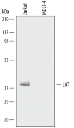
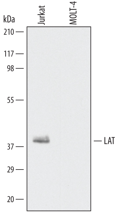
- Detection of Human LAT by Western Blot. Western blot shows lysates of Jurkat human acute T cell leukemia cell line and MOLT-4 human acute lymphoblastic leukemia cell line. PVDF Membrane was probed with 0.1 µg/mL of Human LAT Monoclonal Antibody (Catalog # MAB63341) followed by HRP-conjugated Anti-Mouse IgG Secondary Antibody (Catalog # HAF007). A specific band was detected for LAT at approximately 40 kDa (as indicated). This experiment was conducted under reducing conditions and using Immunoblot Buffer Group 1.
Supportive validation
- Submitted by
- Novus Biologicals (provider)
- Main image
- Experimental details
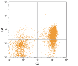
- Detection of LAT in Human PBMCs by Flow Cytometry. Human peripheral blood lymphocytes were stained with Human LAT Monoclonal Antibody (Catalog # MAB63341) followed by Allophycocyanin-conjugated Anti-Mouse IgG Secondary Antibody (Catalog # F0101B) and Human CD3 epsilon PE-conjugated Monoclonal Antibody (Catalog # FAB100P). Quadrant markers were set based on control antibody staining (Catalog # MAB0041).
