 Explore
 Validate
 Learn
Learn Western blot
 Immunoprecipitation
Antibody data
- Antibody Data
- Antigen structure
- References [0]
- Comments [0]
- Validations
- Western blot [2]
- Flow cytometry [4]
Submit
Validation data
Reference
Comment
Report error
- Product number
- MA1-19307 - Provider product page

- Provider
- Invitrogen Antibodies
- Product name
- LAT Monoclonal Antibody (LAT-01)
- Antibody type
- Monoclonal
- Antigen
- Synthetic peptide
- Description
- This antibody reacts with an intracellular epitope of LAT, a 36-38 kDa transmembrane adaptor protein expressed by T cells, pre-B cells, NK cells, mast cells and platelets. This antibody will not cross-react with mouse.
- Reactivity
- Human
- Host
- Mouse
- Isotype
- IgG
- Antibody clone number
- LAT-01
- Vial size
- 100 µg
- Concentration
- 1 mg/mL
- Storage
- 4° C, do not freeze
No comments: Submit comment
Supportive validation
- Submitted by
- Invitrogen Antibodies (provider)
- Main image

- Experimental details
- Western blotting analysis of human LAT using mouse monoclonal antibody LAT-01 in lysates of Jurkat cells (positive) and Raji cells (negative control) under non-reducing and reducing conditions. Nitrocellulose membrane was probed with 2µg/mL of mouse anti-LAT Monoclonal antibody (Product # MA1-19307) followed by IRDye800-conjugated anti-mouse secondary antibody. A specific band was detected for LAT at approximately 38kDa.

- Submitted by
- Invitrogen Antibodies (provider)
- Main image

- Experimental details
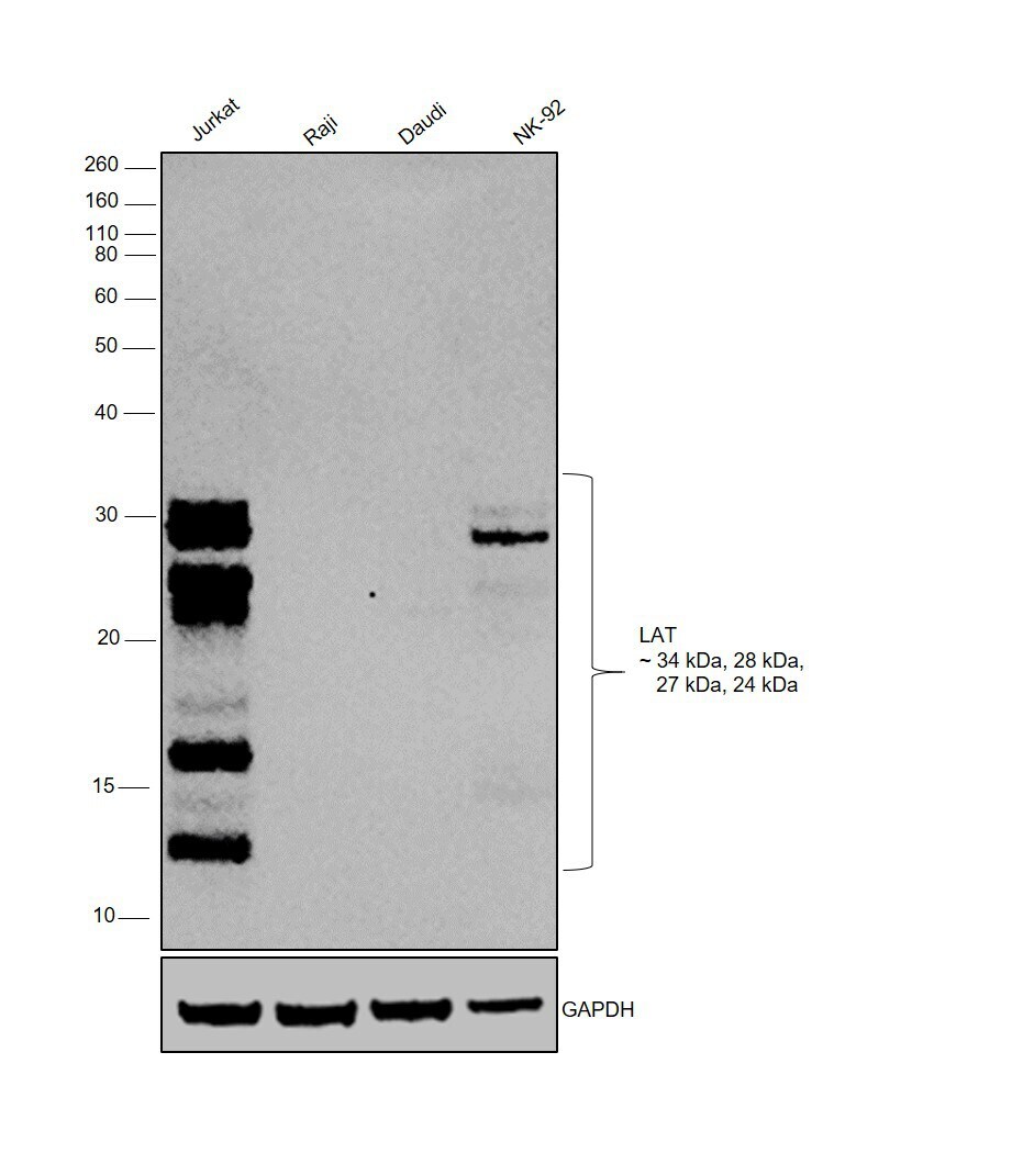
- Western blot was performed using Anti-LAT Monoclonal Antibody (LAT-01)(Product # MA1-19307) and a 34kDa, 28kDa, 27kDa and 24kDa band corresponding to LAT was observed across in T cell line, Jurkat, and Natural Killer cells, NK-92, but not in B-cells (Raji and Daudi). Membrane enriched extracts (30 µg lysate) of Jurkat (Lane 1), Raji (Lane 2), Daudi (Lane 3), NK-92 (Lane 4) were electrophoresed using NuPAGE™ 12% Bis-Tris Protein Gel (Product # NP0341BOX). Resolved proteins were then transferred onto a Nitrocellulose membrane (Product # IB23001) by iBlot® 2 Dry Blotting System (Product # IB21001). The blot was probed with the primary antibody (1:1250) and detected by chemiluminescence with Goat anti-Mouse IgG (H+L) Superclonal™ Recombinant Secondary Antibody, HRP (Product # A28177,1:8000) using the iBright FL 1000 (Product # A32752). Chemiluminescent detection was performed using Novex® ECL Chemiluminescent Substrate Reagent Kit (Product # WP20005).Linker for T cell activation (LAT) is preferentially expressed in higher levels in T-cells and Natural Killer cells for its development and proliferation. Additional proteolytically cleaved products, reported at several molecular weights >13kDa, are detected in Jurkat and NK-cells in comparison to the B-cell lines tested. [doi:10.3389/fimmu.2018.00115, 10.1042/BJ20121135]
Supportive validation
- Submitted by
- Invitrogen Antibodies (provider)
- Main image

- Experimental details
- Flow cytometry analysis of LAT using a monoclonal antibody (Product # MA1-19307).

- Submitted by
- Invitrogen Antibodies (provider)
- Main image

- Experimental details
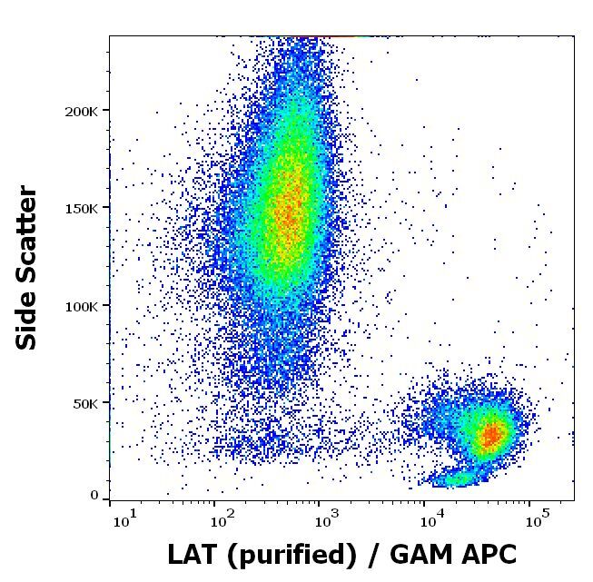
- Flow cytometry intracellular staining pattern of human peripheral whole blood using anti-LAT (LAT-01) purified Monoclonal antibody (Product # MA1-19307) (concentration in sample 1 µg/mL, GAM APC).
- Submitted by
- Invitrogen Antibodies (provider)
- Main image

- Experimental details
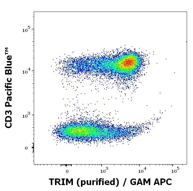
- Flow cytometry multicolor intracellular staining of human peripheral whole blood stained using anti-LAT (LAT-01) purified Monoclonal antibody (Product # MA1-19307) (concentration in sample 1 µg/mL, GAM APC) and anti-human CD3 (UCHT1) Pacific Blue™ using a dilution of 20 µL reagent/100 µL of peripheral whole blood.
- Submitted by
- Invitrogen Antibodies (provider)
- Main image
- Experimental details
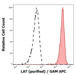
- Separation of human CD3 positive LAT positive lymphocytes (red-filled) from neutrophil granulocytes (black-dashed) in flow cytometry analysis (intracellular staining) of peripheral whole blood stained using anti-LAT (LAT-01) purified Monoclonal antibody (Product # MA1-19307) (concentration in sample 1 µg/mL, GAM APC).
