Explore
 Validate
 Learn
Learn Western blot
 Immunocytochemistry
Antibody data
- Antibody Data
- Antigen structure
- References [1]
- Comments [0]
- Validations
- Immunocytochemistry [1]
- Other assay [2]
Submit
Validation data
Reference
Comment
Report error
- Product number
- PA5-31291 - Provider product page

- Provider
- Invitrogen Antibodies
- Product name
- TRIM10 Polyclonal Antibody
- Antibody type
- Polyclonal
- Antigen
- Recombinant full-length protein
- Description
- Recommended positive controls: NT2D1, IMR32, U87-MG, MCF-7. Predicted reactivity: Rhesus Monkey (95%), Chimpanzee (96%). Store product as a concentrated solution. Centrifuge briefly prior to opening the vial.
- Reactivity
- Human
- Host
- Rabbit
- Isotype
- IgG
- Vial size
- 100 μL
- Concentration
- 0.98 mg/mL
- Storage
- Store at 4°C short term. For long term storage, store at -20°C, avoiding freeze/thaw cycles.
Submitted references TRIM10 binds to IFN-α/β receptor 1 to negatively regulate type I IFN signal transduction.
Guo M, Cao W, Chen S, Tian R, Wang L, Liu Q, Zhang L, Wang Z, Zhao M, Lu Q, Zhu H
European journal of immunology 2021 Jul;51(7):1762-1773
European journal of immunology 2021 Jul;51(7):1762-1773
No comments: Submit comment
Supportive validation
- Submitted by
- Invitrogen Antibodies (provider)
- Main image
- Experimental details
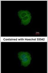
- Immunofluorescent analysis of TRIM10 in methanol-fixed MCF-7 cells using a TRIM10 polyclonal antibody (Product # PA5-31291) at a 1:200 dilution.

Supportive validation
- Submitted by
- Invitrogen Antibodies (provider)
- Main image

- Experimental details
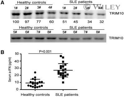
- 1 Figure Decreased serum TRIM10 in patients with systemic lupus erythematosus (SLE). (A) Serum samples of patients and healthy subjects were diluted in phosphate buffer saline (PBS; 1:5). Protein concentration was determined by BCA method. Western blot analysis of serum TRIM10 protein (10 mug) in patients with SLE ( n = 8) and healthy donors ( n = 8) was performed. Raw data for the western blots are shown in Supporting Information Fig. S1A. (B) The level of IFN-alpha in the sera of SLE patients ( n = 20) and healthy donors ( n = 20) was examined by ELISA. (A) The data were shown one representative experiments of three independent experiments with eight donors per experiment. (B) Two-sided Student's t tests were performed to analyze the results. Bars indicated mean and SD of 20 biological replicates, and showed one representative experiment of three independent experiments with all the 20 donors per experiment.
- Submitted by
- Invitrogen Antibodies (provider)
- Main image

- Experimental details
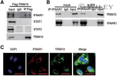
- 4 Figure TRIM10 interacts with IFNAR1. (A) 293T cells were transfected with pFlag-TRIM10. The cells were collected and subjected to the Co-IP assay. Flag-TRIM10 was detected by Co-IP and immunoblotting with the indicated antibodies. Raw data for the western blots were shown in Supporting Information Fig. S4A. (B) HLCZ01 cells were treated with IFN-alpha (200 U/mL) for 15 min. Cell lysates were subjected to IP with control IgG or anti-IFNAR1 antibody. The immunoprecipitants were then probed with anti-TRIM10, anti-IFNAR1 antibody. Raw data for the western blots were shown in Supporting Information Fig. S4B. (C) The subcellular localization of endogenous TRIM10 (green) and IFNAR1 (red) in Huh7 cells was visualized using immunofluorescence with anti-TRIM10 and anti-IFNAR1 antibodies. Cell nuclei were counterstained by DAPI (blue), and observed under confocal microscopy (40x). Scale bars, 50 um. Experiments were independently repeated two (A, B) or three (C) times, with three biological replicates per experiment. Raw data for the western blots were stated as Supporting Information Fig. 4S.