 Explore
 Validate
 Learn
Learn Western blot
 ELISA
ELISA Immunocytochemistry
Antibody data
- Antibody Data
- Antigen structure
- References [0]
- Comments [0]
- Validations
- Immunocytochemistry [2]
- Immunohistochemistry [12]
- Flow cytometry [4]
Submit
Validation data
Reference
Comment
Report error
- Product number
- PA5-114392 - Provider product page

- Provider
- Invitrogen Antibodies
- Product name
- PHF21A Polyclonal Antibody
- Antibody type
- Polyclonal
- Antigen
- Recombinant full-length protein
- Description
- Reconstitute with 0.2 mL of distilled water to yield a concentration of 500 µg/mL. Positive Control - WB: human A549 whole cell, rat brain tissue, mouse brain tissue. IHC: human Ovarian cancer tissue, human oesophagus squama cancer tissue, human tonsil tissue, human endometrial carcinoma tissue, human gastric cancer tissue, human intestinal cancer tissue, human lung cancer tissue, human placenta tissue, human sarcoma tissue, human glioma tissue. Flow: HeLa cell, SiHa cell.|Store at -20°C for one year from date of receipt. After reconstitution, at 4°C for one month. It can also be aliquotted and stored frozen at -20°C for six months. Avoid repeated freeze-thaw cycles.
- Reactivity
- Human, Mouse, Rat
- Host
- Rabbit
- Isotype
- IgG
- Vial size
- 100 μg
- Concentration
- 500 μg/mL
- Storage
- -20°C
No comments: Submit comment
Supportive validation
- Submitted by
- Invitrogen Antibodies (provider)
- Main image

- Experimental details
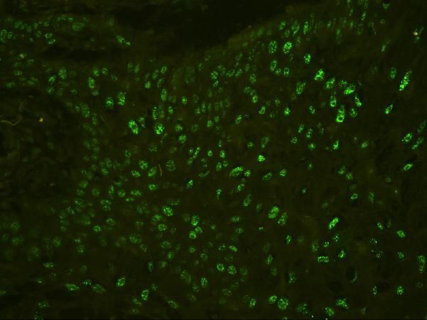
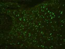
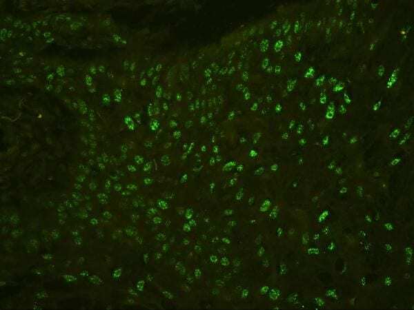
- Immunocytochemistry analysis of PHF21A in human esophagus squama cancer tissues. Antigen retrieval was performed with citrate buffer (pH6, 20 min). Samples were blocked with 10% goat serum and incubated in PHF21A polyclonal antibody (Product # PA5-114392) at a dilution of 1 µg/mL (overnight, 4°C), followed by Biotin conjugated goat anti-rabbit IgG (30 min, 37°C) and DAPI at a dilution of 1:100.
- Submitted by
- Invitrogen Antibodies (provider)
- Main image
- Experimental details
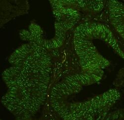
- Immunocytochemistry analysis of PHF21A in human intestinal cancer tissues. Antigen retrieval was performed with citrate buffer (pH6, 20 min). Samples were blocked with 10% goat serum and incubated in PHF21A polyclonal antibody (Product # PA5-114392) at a dilution of 1 µg/mL (overnight, 4°C), followed by Biotin conjugated goat anti-rabbit IgG (30 min, 37°C) and DAPI at a dilution of 1:100.
Supportive validation
- Submitted by
- Invitrogen Antibodies (provider)
- Main image
- Experimental details
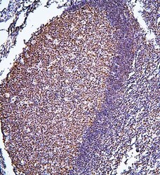
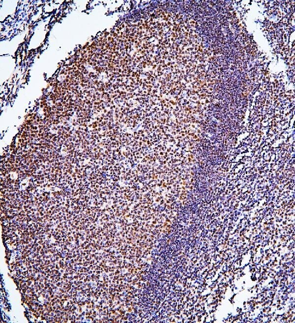
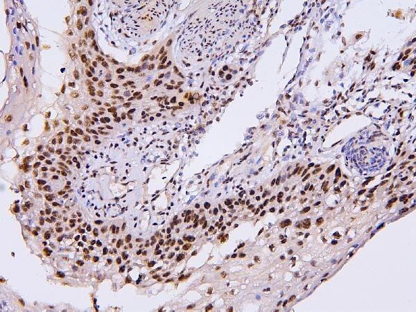
- Immunohistochemistry analysis of PHF21A in paraffin-embedded human tonsil tissues. Antigen retrieval was performed with citrate buffer (pH6, 20 min). Samples were blocked with 10% goat serum and incubated in PHF21A polyclonal antibody (Product # PA5-114392) at a dilution of 1 µg/mL (overnight, 4°C), followed by biotinylated goat anti-rabbit IgG (30 min, 37°C) and Strepavidin-Biotin-Complex (SABC) with DAB.
- Submitted by
- Invitrogen Antibodies (provider)
- Main image
- Experimental details
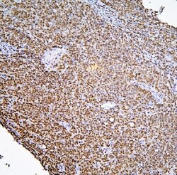
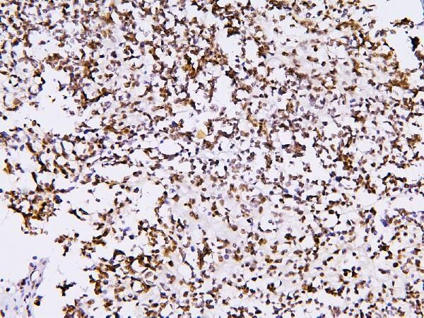
- Immunohistochemistry analysis of PHF21A in paraffin-embedded human sarcoma tissues. Antigen retrieval was performed with citrate buffer (pH6, 20 min). Samples were blocked with 10% goat serum and incubated in PHF21A polyclonal antibody (Product # PA5-114392) at a dilution of 1 µg/mL (overnight, 4°C), followed by biotinylated goat anti-rabbit IgG (30 min, 37°C) and Strepavidin-Biotin-Complex (SABC) with DAB.
- Submitted by
- Invitrogen Antibodies (provider)
- Main image
- Experimental details
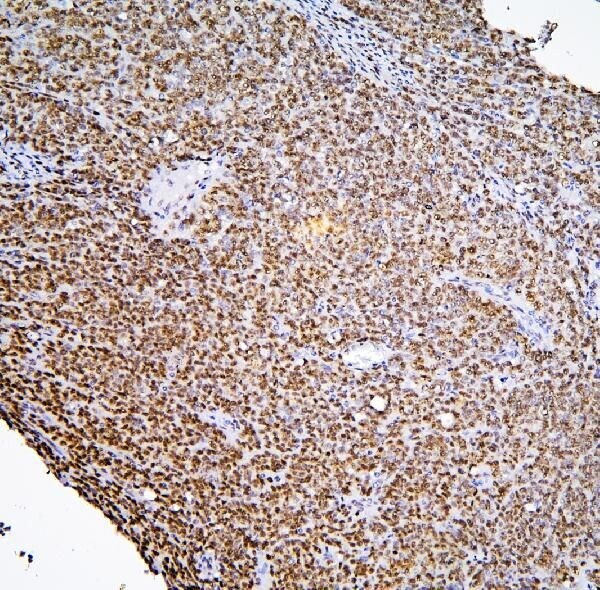
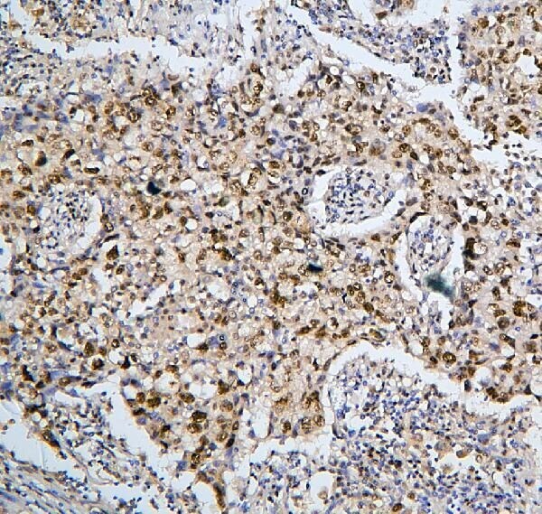
- Immunohistochemistry analysis of PHF21A in paraffin-embedded human Ovarian cancer tissues. Antigen retrieval was performed with citrate buffer (pH6, 20 min). Samples were blocked with 10% goat serum and incubated in PHF21A polyclonal antibody (Product # PA5-114392) at a dilution of 1 µg/mL (overnight, 4°C), followed by biotinylated goat anti-rabbit IgG (30 min, 37°C) and Strepavidin-Biotin-Complex (SABC) with DAB.
- Submitted by
- Invitrogen Antibodies (provider)
- Main image
- Experimental details
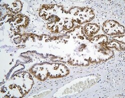
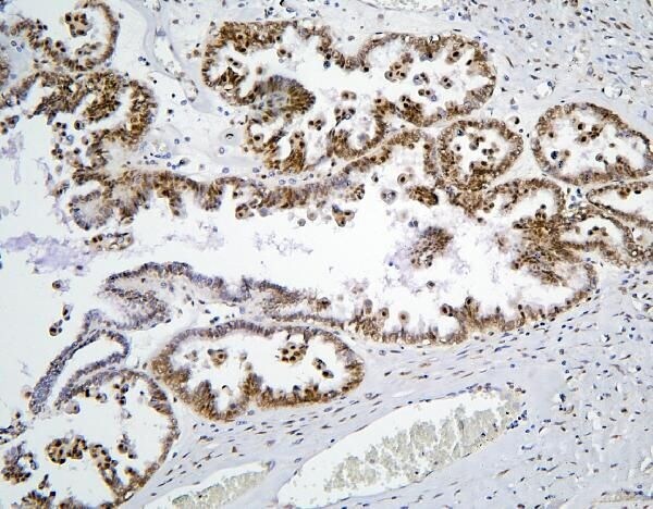
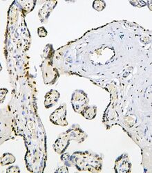
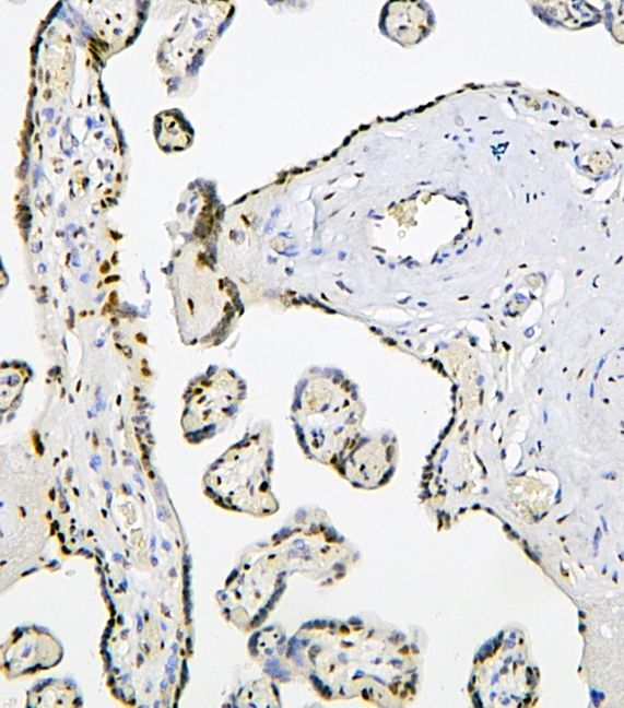
- Immunohistochemistry analysis of PHF21A in paraffin-embedded human placenta tissues. Antigen retrieval was performed with citrate buffer (pH6, 20 min). Samples were blocked with 10% goat serum and incubated in PHF21A polyclonal antibody (Product # PA5-114392) at a dilution of 1 µg/mL (overnight, 4°C), followed by biotinylated goat anti-rabbit IgG (30 min, 37°C) and Strepavidin-Biotin-Complex (SABC) with DAB.
- Submitted by
- Invitrogen Antibodies (provider)
- Main image
- Experimental details
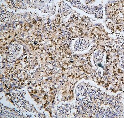
- Immunohistochemistry analysis of PHF21A in paraffin-embedded human lung cancer tissues. Antigen retrieval was performed with citrate buffer (pH6, 20 min). Samples were blocked with 10% goat serum and incubated in PHF21A polyclonal antibody (Product # PA5-114392) at a dilution of 1 µg/mL (overnight, 4°C), followed by biotinylated goat anti-rabbit IgG (30 min, 37°C) and Strepavidin-Biotin-Complex (SABC) with DAB.
- Submitted by
- Invitrogen Antibodies (provider)
- Main image

- Experimental details
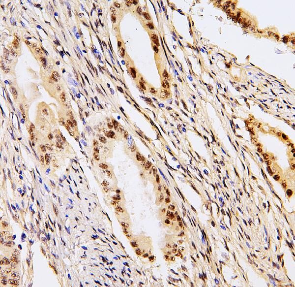
- Immunohistochemistry analysis of PHF21A in paraffin-embedded human intestinal cancer tissues. Antigen retrieval was performed with citrate buffer (pH6, 20 min). Samples were blocked with 10% goat serum and incubated in PHF21A polyclonal antibody (Product # PA5-114392) at a dilution of 1 µg/mL (overnight, 4°C), followed by biotinylated goat anti-rabbit IgG (30 min, 37°C) and Strepavidin-Biotin-Complex (SABC) with DAB.
- Submitted by
- Invitrogen Antibodies (provider)
- Main image
- Experimental details
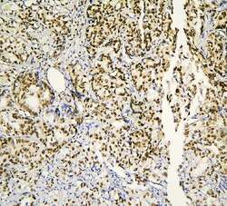
- Immunohistochemistry analysis of PHF21A in paraffin-embedded human gastric cancer tissues. Antigen retrieval was performed with citrate buffer (pH6, 20 min). Samples were blocked with 10% goat serum and incubated in PHF21A polyclonal antibody (Product # PA5-114392) at a dilution of 1 µg/mL (overnight, 4°C), followed by biotinylated goat anti-rabbit IgG (30 min, 37°C) and Strepavidin-Biotin-Complex (SABC) with DAB.
- Submitted by
- Invitrogen Antibodies (provider)
- Main image

- Experimental details
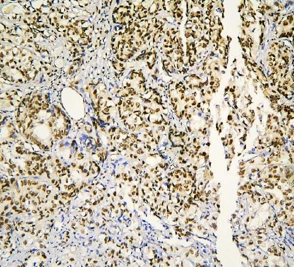
- Immunohistochemistry analysis of PHF21A in paraffin-embedded human endometrial carcinoma tissues. Antigen retrieval was performed with citrate buffer (pH6, 20 min). Samples were blocked with 10% goat serum and incubated in PHF21A polyclonal antibody (Product # PA5-114392) at a dilution of 1 µg/mL (overnight, 4°C), followed by biotinylated goat anti-rabbit IgG (30 min, 37°C) and Strepavidin-Biotin-Complex (SABC) with DAB.
- Submitted by
- Invitrogen Antibodies (provider)
- Main image

- Experimental details
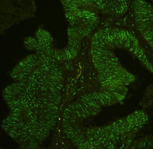
- Immunocytochemistry analysis of PHF21A in paraffin-embedded human intestinal cancer tissues. Antigen retrieval was performed with citrate buffer (pH6, 20 min). Samples were blocked with 10% goat serum and incubated in PHF21A polyclonal antibody (Product # PA5-114392) at a dilution of 1 µg/mL (overnight, 4°C), followed by Biotin conjugated goat anti-rabbit IgG (30 min, 37°C) and DAPI at a dilution of 1:100.

- Submitted by
- Invitrogen Antibodies (provider)
- Main image

- Experimental details
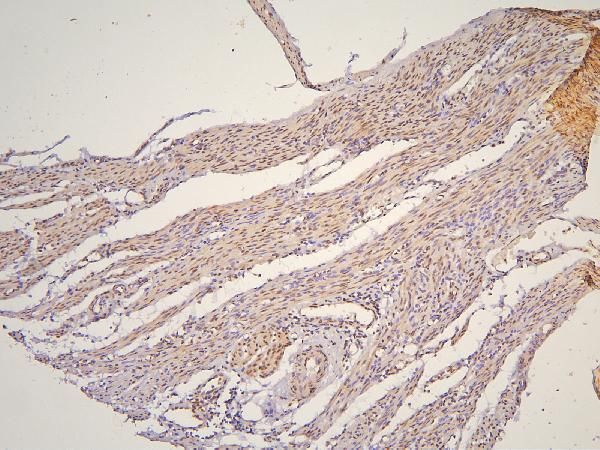
- Immunohistochemistry analysis of PHF21A in paraffin-embedded human esophagus squama cancer tissues. Antigen retrieval was performed with citrate buffer (pH6, 20 min). Samples were blocked with 10% goat serum and incubated in PHF21A polyclonal antibody (Product # PA5-114392) at a dilution of 1 µg/mL (overnight, 4°C), followed by biotinylated goat anti-rabbit IgG (30 min, 37°C) and Strepavidin-Biotin-Complex (SABC) with DAB.
- Submitted by
- Invitrogen Antibodies (provider)
- Main image

- Experimental details
- Immunohistochemistry analysis of PHF21A in paraffin-embedded human glioma tissues. Antigen retrieval was performed with citrate buffer (pH6, 20 min). Samples were blocked with 10% goat serum and incubated in PHF21A polyclonal antibody (Product # PA5-114392) at a dilution of 1 µg/mL (overnight, 4°C), followed by biotinylated goat anti-rabbit IgG (30 min, 37°C) and Strepavidin-Biotin-Complex (SABC) with DAB.
- Submitted by
- Invitrogen Antibodies (provider)
- Main image
- Experimental details
- Immunocytochemistry analysis of PHF21A in paraffin-embedded human esophagus squama cancer tissues. Antigen retrieval was performed with citrate buffer (pH6, 20 min). Samples were blocked with 10% goat serum and incubated in PHF21A polyclonal antibody (Product # PA5-114392) at a dilution of 1 µg/mL (overnight, 4°C), followed by Biotin conjugated goat anti-rabbit IgG (30 min, 37°C) and DAPI at a dilution of 1:100.
Supportive validation
- Submitted by
- Invitrogen Antibodies (provider)
- Main image
- Experimental details
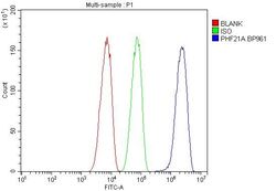
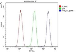
- Flow cytometry analysis of PHF21A in SiHa cells. Samples were blocked with 10% normal goat serum and incubated in PHF21A polyclonal antibody (Product # PA5-114392) at a dilution of 1 µg/1x10^6 cells (30 min, 20°C), followed by DyLight 488 conjugated goat anti-rabbit IgG (30 min, 20°C) with a dilution of 5-10 µg/1x10^6 cells (30 min, 20°C). Isotype control antibody (Green line) was rabbit IgG (1 µg/1x10^6) and unlabeled sample (Red line).
- Submitted by
- Invitrogen Antibodies (provider)
- Main image

- Experimental details
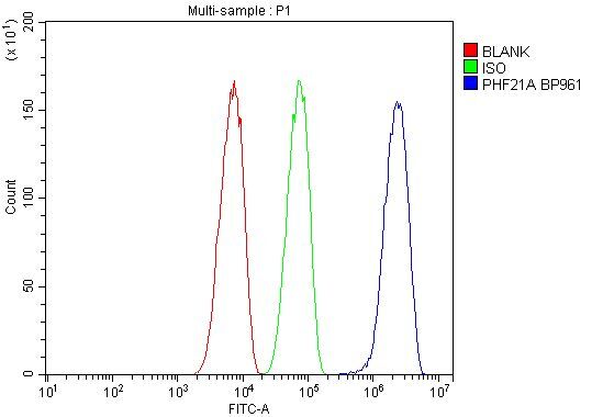
- Flow cytometry analysis of PHF21A in HeLA cells. Samples were blocked with 10% normal goat serum and incubated in PHF21A polyclonal antibody (Product # PA5-114392) at a dilution of 1 µg/1x10^6 cells (30 min, 20°C), followed by DyLight 488 conjugated goat anti-rabbit IgG (30 min, 20°C) with a dilution of 5-10 µg/1x10^6 cells (30 min, 20°C). Isotype control antibody (Green line) was rabbit IgG (1 µg/1x10^6) and unlabeled sample (Red line).
- Submitted by
- Invitrogen Antibodies (provider)
- Main image
- Experimental details
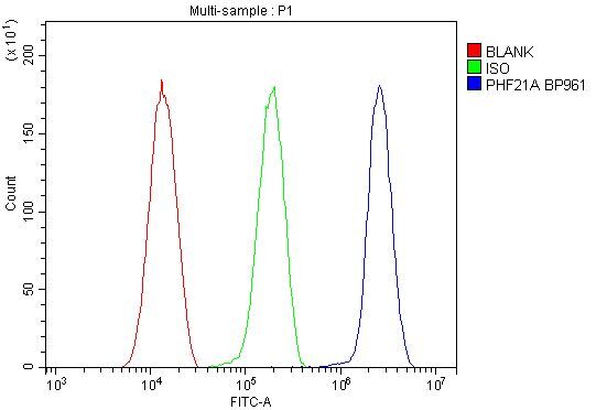
- Flow cytometry analysis of PHF21A in HeLA cells. Samples were blocked with 10% normal goat serum and incubated in PHF21A polyclonal antibody (Product # PA5-114392) at a dilution of 1 µg/1x10^6 cells (30 min, 20°C), followed by DyLight 488 conjugated goat anti-rabbit IgG (30 min, 20°C) with a dilution of 5-10 µg/1x10^6 cells (30 min, 20°C). Isotype control antibody (Green line) was rabbit IgG (1 µg/1x10^6) and unlabeled sample (Red line).

- Submitted by
- Invitrogen Antibodies (provider)
- Main image

- Experimental details
- Flow cytometry analysis of PHF21A in SiHa cells. Samples were blocked with 10% normal goat serum and incubated in PHF21A polyclonal antibody (Product # PA5-114392) at a dilution of 1 µg/1x10^6 cells (30 min, 20°C), followed by DyLight 488 conjugated goat anti-rabbit IgG (30 min, 20°C) with a dilution of 5-10 µg/1x10^6 cells (30 min, 20°C). Isotype control antibody (Green line) was rabbit IgG (1 µg/1x10^6) and unlabeled sample (Red line).
