Explore
 Validate
 Learn
Learn Western blot
 Immunocytochemistry
Antibody data
- Antibody Data
- Antigen structure
- References [11]
- Comments [0]
- Validations
- Western blot [2]
- Immunohistochemistry [1]
- Flow cytometry [2]
Submit
Validation data
Reference
Comment
Report error
- Product number
- AF1172 - Provider product page

- Provider
- Novus Biologicals
- Product name
- Goat Polyclonal ALCAM/CD166 Antibody
- Antibody type
- Polyclonal
- Description
- Antigen Affinity-purified. Detects human, mouse and rat ALCAM/CD166 in Western blots. In direct ELISAs, less than 10% cross-reactivity with rhBCAM, recombinant mouse (rm) OCAM, and rmMAdCAM-1 is observed. Detects canine ALCAM/CD166 in flow cytometry.
- Reactivity
- Human, Mouse, Rat, Canine
- Host
- Goat
- Conjugate
- Unconjugated
- Isotype
- IgG
- Vial size
- 100 ug
- Concentration
- LYOPH
- Storage
- Use a manual defrost freezer and avoid repeated freeze-thaw cycles. 12 months from date of receipt, -20 to -70 degreesC as supplied. 1 month, 2 to 8 degreesC under sterile conditions after reconstitution. 6 months, -20 to -70 degreesC under sterile conditions after reconstitution.
Submitted references Axonal Growth of Midbrain Dopamine Neurons is Modulated by the Cell Adhesion Molecule ALCAM Through Trans-Heterophilic Interactions with L1cam, Chl1, and Semaphorins.
Tbr1 instructs laminar patterning of retinal ganglion cell dendrites.
Multicolor quantitative confocal imaging cytometry.
Dual role of ALCAM in neuroinflammation and blood-brain barrier homeostasis.
Expression of the immunoglobulin superfamily cell adhesion molecules in the developing spinal cord and dorsal root ganglion.
Transcriptome analysis reveals transmembrane targets on transplantable midbrain dopamine progenitors.
Dbx1 triggers crucial molecular programs required for midline crossing by midbrain commissural axons.
Ionizing irradiation induces acute haematopoietic syndrome and gastrointestinal syndrome independently in mice.
Cocaine hijacks σ1 receptor to initiate induction of activated leukocyte cell adhesion molecule: implication for increased monocyte adhesion and migration in the CNS.
Molecular mechanisms controlling midline crossing by precerebellar neurons.
Inhibition of p38 MAPK signaling in chondrocyte cultures results in enhanced osteogenic differentiation of perichondral cells.
Bye CR, Rytova V, Alsanie WF, Parish CL, Thompson LH
The Journal of neuroscience : the official journal of the Society for Neuroscience 2019 Aug 21;39(34):6656-6667
The Journal of neuroscience : the official journal of the Society for Neuroscience 2019 Aug 21;39(34):6656-6667
Tbr1 instructs laminar patterning of retinal ganglion cell dendrites.
Liu J, Reggiani JDS, Laboulaye MA, Pandey S, Chen B, Rubenstein JLR, Krishnaswamy A, Sanes JR
Nature neuroscience 2018 May;21(5):659-670
Nature neuroscience 2018 May;21(5):659-670
Multicolor quantitative confocal imaging cytometry.
Coutu DL, Kokkaliaris KD, Kunz L, Schroeder T
Nature methods 2018 Jan;15(1):39-46
Nature methods 2018 Jan;15(1):39-46
Dual role of ALCAM in neuroinflammation and blood-brain barrier homeostasis.
Lécuyer MA, Saint-Laurent O, Bourbonnière L, Larouche S, Larochelle C, Michel L, Charabati M, Abadier M, Zandee S, Haghayegh Jahromi N, Gowing E, Pittet C, Lyck R, Engelhardt B, Prat A
Proceedings of the National Academy of Sciences of the United States of America 2017 Jan 24;114(4):E524-E533
Proceedings of the National Academy of Sciences of the United States of America 2017 Jan 24;114(4):E524-E533
Expression of the immunoglobulin superfamily cell adhesion molecules in the developing spinal cord and dorsal root ganglion.
Gu Z, Imai F, Kim IJ, Fujita H, Katayama Ki, Mori K, Yoshihara Y, Yoshida Y
PloS one 2015;10(3):e0121550
PloS one 2015;10(3):e0121550
Transcriptome analysis reveals transmembrane targets on transplantable midbrain dopamine progenitors.
Bye CR, Jönsson ME, Björklund A, Parish CL, Thompson LH
Proceedings of the National Academy of Sciences of the United States of America 2015 Apr 14;112(15):E1946-55
Proceedings of the National Academy of Sciences of the United States of America 2015 Apr 14;112(15):E1946-55
Dbx1 triggers crucial molecular programs required for midline crossing by midbrain commissural axons.
Inamata Y, Shirasaki R
Development (Cambridge, England) 2014 Mar;141(6):1260-71
Development (Cambridge, England) 2014 Mar;141(6):1260-71
Ionizing irradiation induces acute haematopoietic syndrome and gastrointestinal syndrome independently in mice.
Leibowitz BJ, Wei L, Zhang L, Ping X, Epperly M, Greenberger J, Cheng T, Yu J
Nature communications 2014 Mar 18;5:3494
Nature communications 2014 Mar 18;5:3494
Cocaine hijacks σ1 receptor to initiate induction of activated leukocyte cell adhesion molecule: implication for increased monocyte adhesion and migration in the CNS.
Yao H, Kim K, Duan M, Hayashi T, Guo M, Morgello S, Prat A, Wang J, Su TP, Buch S
The Journal of neuroscience : the official journal of the Society for Neuroscience 2011 Apr 20;31(16):5942-55
The Journal of neuroscience : the official journal of the Society for Neuroscience 2011 Apr 20;31(16):5942-55
Molecular mechanisms controlling midline crossing by precerebellar neurons.
Di Meglio T, Nguyen-Ba-Charvet KT, Tessier-Lavigne M, Sotelo C, Chédotal A
The Journal of neuroscience : the official journal of the Society for Neuroscience 2008 Jun 18;28(25):6285-94
The Journal of neuroscience : the official journal of the Society for Neuroscience 2008 Jun 18;28(25):6285-94
Inhibition of p38 MAPK signaling in chondrocyte cultures results in enhanced osteogenic differentiation of perichondral cells.
Stanton LA, Beier F
Experimental cell research 2007 Jan 1;313(1):146-55
Experimental cell research 2007 Jan 1;313(1):146-55
No comments: Submit comment
Supportive validation
- Submitted by
- Novus Biologicals (provider)
- Main image
- Experimental details
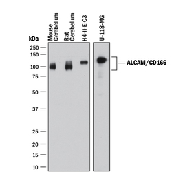
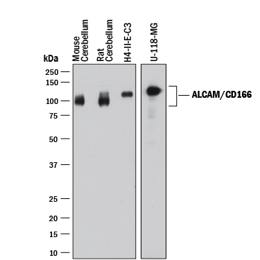
- Detection of Human, Mouse, and Rat ALCAM/CD166 by Western Blot. Western blot shows lysates of mouse brain (cerebellumn) tissue, rat brain (cerebellum) tissue, H4-II-E-C3 rat hepatoma cell line, and U-118-MG human glioblastoma/astrocytoma cell line. PVDF membrane was probed with 0.2 µg/mL of Goat Anti-Mouse/Rat/Canine ALCAM/CD166 Antigen Affinity-purified Polyclonal Antibody (Catalog # AF1172) followed by HRP-conjugated Anti-Goat IgG Secondary Antibody (Catalog # HAF017). A specific band was detected for ALCAM/CD166 at approximately 90-120 kDa (as indicated). This experiment was conducted under reducing conditions and using Immunoblot Buffer Group 1.
- Submitted by
- Novus Biologicals (provider)
- Main image

- Experimental details
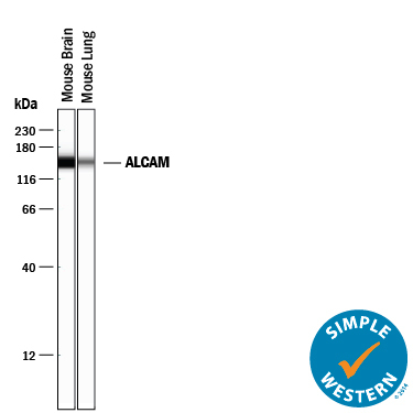
- Detection of Mouse ALCAM/CD166 by Simple WesternTM. Simple Western lane view shows lysates of mouse brain tissue and mouse lung tissue loaded at 0.2 mg/mL. A specific band was detected for ALCAM/CD166 at approximately 149 kDa (as indicated) using 12.5 µg/mL of Goat Anti-Mouse/Rat/Canine ALCAM/CD166 Antigen Affinity-purified Polyclonal Antibody (Catalog # AF1172) followed by 1:50 dilution of HRP-conjugated Anti-Goat IgG Secondary Antibody (Catalog # HAF109). This experiment was conducted under reducing conditions and using the 12-230 kDa separation system.
Supportive validation
- Submitted by
- Novus Biologicals (provider)
- Main image

- Experimental details
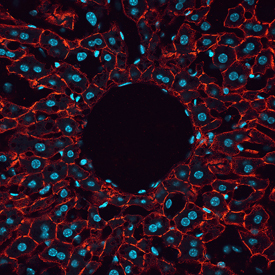
- ALCAM/CD166 in Mouse Liver. ALCAM/CD166 was detected in perfusion fixed frozen sections of mouse liver using Goat Anti-Mouse/Rat/Canine ALCAM/CD166 Antigen Affinity-purified Polyclonal Antibody (Catalog # AF1172) at 1.7 µg/mL overnight at 4 °C. Tissue was stained using the NorthernLights™ 557-conjugated Anti-Goat IgG Secondary Antibody (red; Catalog # NL001) and counterstained with DAPI (blue). View our protocol for Fluorescent IHC Staining of Frozen Tissue Sections.
Supportive validation
- Submitted by
- Novus Biologicals (provider)
- Main image
- Experimental details
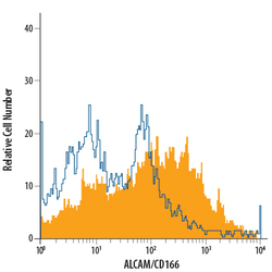
- Detection of ALCAM/CD166 in Canine PBMCs by Flow Cytometry. Canine peripheral blood mononuclear cells (PBMCs) treated with PMA and Calcium Ionomycin for 24 hours were stained with Goat Anti-Mouse/Rat/Canine ALCAM/CD166 Antigen Affinity-purified Polyclonal Antibody (Catalog # AF1172, filled histogram) or isotype control antibody (Catalog # AB-108-C, open histogram), followed by Phycoerythrin-conjugated Anti-Goat IgG Secondary Antibody (Catalog # F0107).
- Submitted by
- Novus Biologicals (provider)
- Main image
- Experimental details
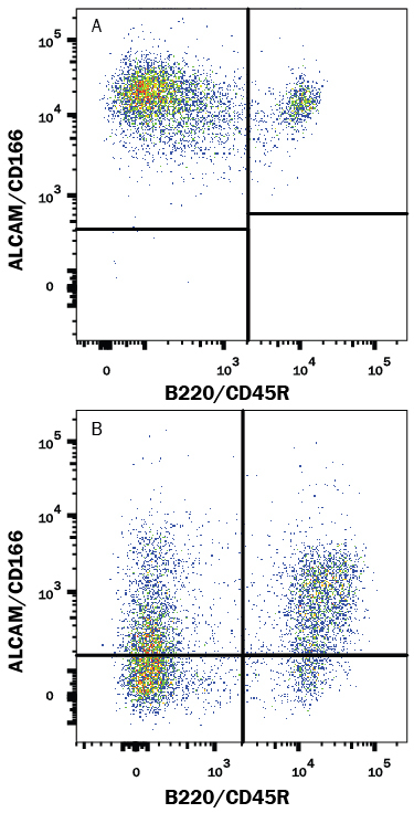
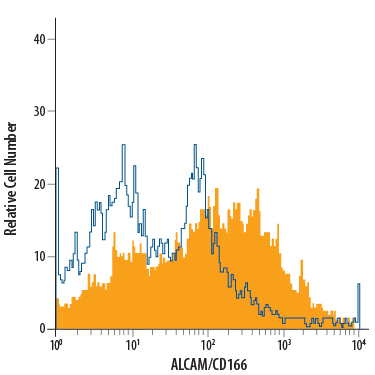
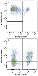
- Detection of ALCAM/CD166 in Mouse Splenocytes by Flow Cytometry. Mouse splenocytes either (A) activated or (B) resting were stained with Goat Anti-Mouse/Rat/Canine ALCAM/CD166 Antigen Affinity-purified Polyclonal Antibody (Catalog # AF1172) followed by Phycoerythrin-conjugated Anti-Goat IgG Secondary Antibody (Catalog # F0107) and Rat Anti-Mouse B220/CD45R APC-conjugated Monoclonal Antibody (Catalog # FAB1217A). Quadrant markers were set based on control antibody staining (Catalog # AB-108-C).