Explore
 Validate
 Learn
Learn Western blot
 ELISA
ELISAAntibody data
- Antibody Data
- Antigen structure
- References [0]
- Comments [0]
- Validations
- Western blot [3]
- Immunocytochemistry [3]
- Immunohistochemistry [4]
- Flow cytometry [2]
Submit
Validation data
Reference
Comment
Report error
- Product number
- MA5-38443 - Provider product page

- Provider
- Invitrogen Antibodies
- Product name
- CD166 Monoclonal Antibody (8E12C7)
- Antibody type
- Monoclonal
- Antigen
- Purifed from natural sources
- Description
- This antibody has been tested in indirect ELISA.
- Reactivity
- Human
- Host
- Mouse
- Isotype
- IgG
- Antibody clone number
- 8E12C7
- Vial size
- 100 μg
- Concentration
- 1 mg/mL
- Storage
- Store at 4°C short term. For long term storage, store at -20°C, avoiding freeze/thaw cycles.
No comments: Submit comment
Supportive validation
- Submitted by
- Invitrogen Antibodies (provider)
- Main image
- Experimental details
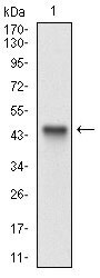
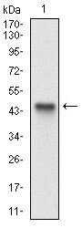
- Western Blot analysis using CD166 Monoclonal Antibody (Product # MA5-38443) against human ALCAM recombinant protein. (Expected MW is 44.9 kDa)
- Submitted by
- Invitrogen Antibodies (provider)
- Main image

- Experimental details
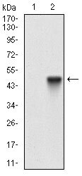
- Western Blot analysis of CD166 (ALCAM) using CD166 Monoclonal Antibody (Product # MA5-38443) in HEK293 (1) and ALCAM (AA: 48-216)-human IgG-Fc transfected HEK293 (2) cell lysate.
- Submitted by
- Invitrogen Antibodies (provider)
- Main image
- Experimental details
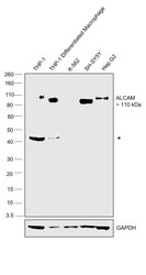
- Western blot was performed using CD166 Monoclonal Antibody (8E12C7) (Product # MA5-38443) and a 110 kDa band corresponding to ALCAM was observed across lanes except for K-562. Whole cell extracts (30 µg lysate) of THP-1 (Lane 1), THP-1 differentiated macrophages (Lane 2), K-562 (Lane 3), SH-SY5Y (Lane 4), Hep G2 (Lane 5) were electrophoresed using NuPAGE™ 4-12% Bis-Tris Protein Gel (Product # NP0321BOX), 10 well. Resolved proteins were then transferred onto a nitrocellulose membrane (Product # IB23001) by iBlot® 2 Dry Blotting System (Product # IB21001). The blot was probed with the primary antibody (1:500) and detected by chemiluminescence with Goat anti-Mouse IgG (H+L) Superclonal™ Recombinant Secondary Antibody, HRP (Product # A28177, 1:10,000) using the iBright™ FL1500 Imaging System (Product # A44115). Chemiluminescentdetection was performed using Novex® ECL Chemiluminescent Substrate Reagent Kit (Product # WP20005). An uncharacterized band (*) at ~ 40 kDa was observed in first two lanes.

Supportive validation
- Submitted by
- Invitrogen Antibodies (provider)
- Main image
- Experimental details
- Immunocytochemistry/Immunofluorescence analysis of CD166 (ALCAM) in HeLa cells using CD166 Monoclonal Antibody (Product # MA5-38443) (green). Blue: DRAQ5 fluorescent DNA dye.

- Submitted by
- Invitrogen Antibodies (provider)
- Main image

- Experimental details
- Immunocytochemistry/Immunofluorescence analysis of CD166 (ALCAM) in HeLa cells using CD166 Monoclonal Antibody (Product # MA5-38443) (green). Blue: DRAQ5 fluorescent DNA dye.

- Submitted by
- Invitrogen Antibodies (provider)
- Main image
- Experimental details
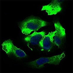
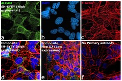
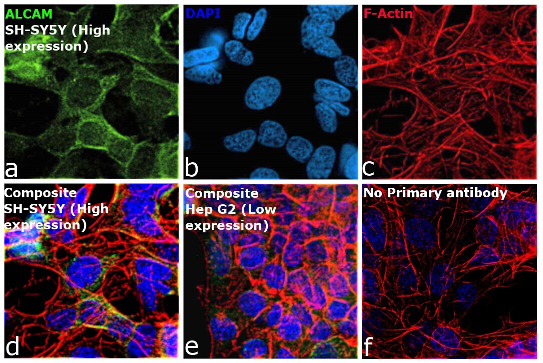
- Immunofluorescence analysis of ALCAM was performed using 70% confluent log phase SH-SY5Y cells. The cells were fixed with 4% paraformaldehyde for 10 minutes, permeabilized with 0.1% Triton™ X-100 for 10 minutes, and blocked with 2% BSA for 45 minutes at room temperature. The cells were labeled with CD166 Monoclonal Antibody (8E12C7) (Product # MA5-38443, 1:250) in 0.1% BSA, incubated at 4 degree celsius overnight and then labeled with Donkey anti-Mouse IgG (H+L) Highly Cross-Adsorbed Secondary Antibody, Alexa Fluor™ Plus 488 (Product # A32766, 1:2500), for 45 minutes at room temperature (Panel a: Green). Nuclei (Panel b: Blue) were stained with ProLong™ Diamond Antifade Mountant with DAPI (Product # P36962). F-actin (Panel c: Red) was stained with Rhodamine Phalloidin (Product # R415, 1:300). Panel d represents the merged image showing plasma membrane and cytosolic localization. Panel e represents Hep G2 cells which show relatively lower levels of the target protein. Panel f represents control cells with no primary antibody to assess background. The images were captured at 60X magnification.
Supportive validation
- Submitted by
- Invitrogen Antibodies (provider)
- Main image
- Experimental details
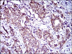
- Immunohistochemistry (Paraffin) analysis of CD166 (ALCAM) in paraffin-embedded cervical cancer tissues using CD166 Monoclonal Antibody (Product # MA5-38443) and DAB staining.
- Submitted by
- Invitrogen Antibodies (provider)
- Main image
- Experimental details
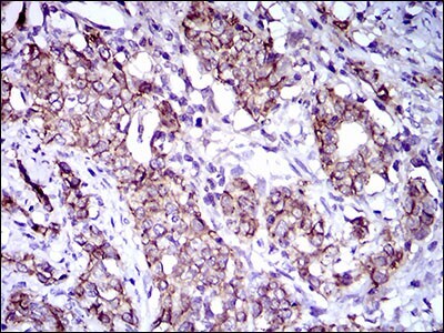
- Immunohistochemistry (Paraffin) analysis of CD166 (ALCAM) in paraffin-embedded bladder cancer tissues using CD166 Monoclonal Antibody (Product # MA5-38443) and DAB staining.

- Submitted by
- Invitrogen Antibodies (provider)
- Main image

- Experimental details
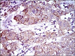
- Immunohistochemistry (Paraffin) analysis of CD166 (ALCAM) in paraffin-embedded cervical cancer tissues using CD166 Monoclonal Antibody (Product # MA5-38443) and DAB staining.

- Submitted by
- Invitrogen Antibodies (provider)
- Main image

- Experimental details
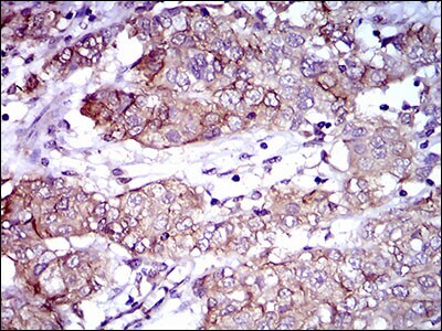
- Immunohistochemistry (Paraffin) analysis of CD166 (ALCAM) in paraffin-embedded bladder cancer tissues using CD166 Monoclonal Antibody (Product # MA5-38443) and DAB staining.
Supportive validation
- Submitted by
- Invitrogen Antibodies (provider)
- Main image

- Experimental details
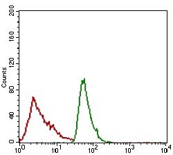
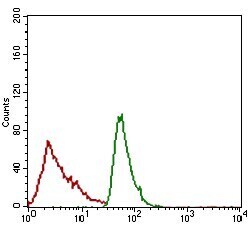
- Flow Cytometry analysis of CD166 (ALCAM) in JURKAT cells using CD166 Monoclonal Antibody (Product # MA5-38443) (green) and negative control (red).

- Submitted by
- Invitrogen Antibodies (provider)
- Main image
- Experimental details
- Flow Cytometry analysis of CD166 (ALCAM) in JURKAT cells using CD166 Monoclonal Antibody (Product # MA5-38443) (green) and negative control (red).