Explore
 Validate
 Learn
Learn Western blot
 Immunocytochemistry
Antibody data
- Antibody Data
- Antigen structure
- References [0]
- Comments [0]
- Validations
- Western blot [4]
- Immunohistochemistry [3]
Submit
Validation data
Reference
Comment
Report error
- Product number
- NBP2-16776 - Provider product page

- Provider
- Novus Biologicals
- Product name
- Rabbit Polyclonal HADH Antibody
- Antibody type
- Polyclonal
- Description
- Immunogen affinity purified.
- Reactivity
- Human, Mouse, Rat
- Host
- Rabbit
- Isotype
- IgG
- Vial size
- 0.1 ml
- Storage
- Aliquot and store at -20C or -80C. Avoid freeze-thaw cycles.
No comments: Submit comment
Supportive validation
- Submitted by
- Novus Biologicals (provider)
- Main image
- Experimental details
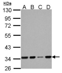
- Western Blot: HADH Antibody [NBP2-16776] - A. 30 ug 293T whole cell lysate/extract B. 30 ug A431 whole cell lysate/extract C. 30 ug HeLa whole cell lysate/extract D. 30 ug HepG2 whole cell lysate/extract 10 % SDS-PAGE

- Submitted by
- Novus Biologicals (provider)
- Main image

- Experimental details
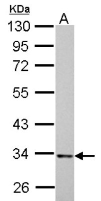
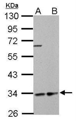
- Western Blot: HADH Antibody [NBP2-16776] - 30 ug PC-12 whole cell lysate/extract 10 % SDS-PAGE HADH antibody dilution: 1:1000.
- Submitted by
- Novus Biologicals (provider)
- Main image
- Experimental details
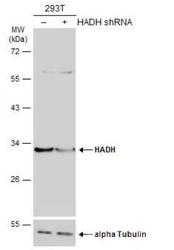
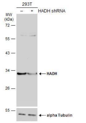
- Western Blot: HADH Antibody [NBP2-16776] - Non-transfected (-) and transfected (+) 293T whole cell extracts (30 ug) were separated by 10% SDS-PAGE, and the membrane was blotted with HADH antibody diluted at 1:1000. HRP-conjugated anti-rabbit IgG antibody was used to detect the primary antibody.
- Submitted by
- Novus Biologicals (provider)
- Main image
- Experimental details
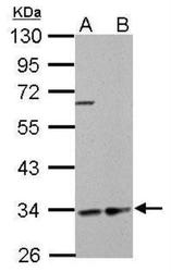
- Western Blot: HADH Antibody [NBP2-16776] - HADH antibody detects HADH protein by Western blot analysis. A. 30 ug GL261 whole cell lysate/extract. B. 30 ug C8D30 whole cell lysate/extract. 10 % SDS-PAGE. HADH antibody dilution: 1:1000.
Supportive validation
- Submitted by
- Novus Biologicals (provider)
- Main image
- Experimental details
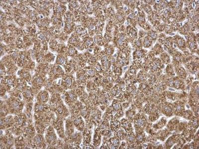
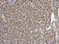
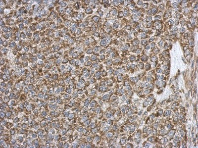
- Immunohistochemistry-Paraffin: HADH Antibody [NBP2-16776] - Immunohistochemical analysis of paraffin-embedded Hela xenograft, using antibody at 1:500 dilution.
- Submitted by
- Novus Biologicals (provider)
- Main image
- Experimental details
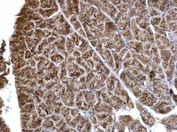
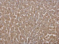
- Immunohistochemistry-Paraffin: HADH Antibody [NBP2-16776] - HADH antibody detects HADH protein at mitochondria on mouse liver by immunohistochemical analysis. Sample: Paraffin-embedded mouse pancreas. HADH antibody dilution: 1:500.
- Submitted by
- Novus Biologicals (provider)
- Main image
- Experimental details
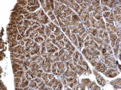
- Immunohistochemistry-Paraffin: HADH Antibody [NBP2-16776] - HADH antibody detects HADH protein at mitochondria on mouse liver by immunohistochemical analysis. Sample: Paraffin-embedded mouse pancreas. HADH antibody dilution: 1:500.