Explore
 Validate
 Learn
Learn Western blot
 Immunoprecipitation
Antibody data
- Antibody Data
- Antigen structure
- References [3]
- Comments [0]
- Validations
- Western blot [2]
- ELISA [1]
- Immunocytochemistry [1]
- Immunohistochemistry [1]
Submit
Validation data
Reference
Comment
Report error
- Product number
- H00003033-M01 - Provider product page

- Provider
- Novus Biologicals
- Proper citation
- Novus Cat#H00003033-M01, RRID:AB_2115573
- Product name
- Mouse Monoclonal HADH Antibody
- Antibody type
- Monoclonal
- Description
- IgG purified. HADHSC - L-3-hydroxyacyl-Coenzyme A dehydrogenase, short chain
- Reactivity
- Human
- Host
- Mouse
- Isotype
- IgG
- Vial size
- 0.1 mg
- Storage
- Aliquot and store at -20C or -80C. Avoid freeze-thaw cycles.
Submitted references Changes in peak fat oxidation in response to different doses of endurance training.
Independent effects of endurance training and weight loss on peak fat oxidation in moderately overweight men: a randomized controlled trial.
BCAT1 promotes cell proliferation through amino acid catabolism in gliomas carrying wild-type IDH1.
Rosenkilde M, Reichkendler MH, Auerbach P, Bonne TC, Sjödin A, Ploug T, Stallknecht BM
Scandinavian journal of medicine & science in sports 2015 Feb;25(1):41-52
Scandinavian journal of medicine & science in sports 2015 Feb;25(1):41-52
Independent effects of endurance training and weight loss on peak fat oxidation in moderately overweight men: a randomized controlled trial.
Nordby P, Rosenkilde M, Ploug T, Westh K, Feigh M, Nielsen NB, Helge JW, Stallknecht B
Journal of applied physiology (Bethesda, Md. : 1985) 2015 Apr 1;118(7):803-10
Journal of applied physiology (Bethesda, Md. : 1985) 2015 Apr 1;118(7):803-10
BCAT1 promotes cell proliferation through amino acid catabolism in gliomas carrying wild-type IDH1.
Tönjes M, Barbus S, Park YJ, Wang W, Schlotter M, Lindroth AM, Pleier SV, Bai AHC, Karra D, Piro RM, Felsberg J, Addington A, Lemke D, Weibrecht I, Hovestadt V, Rolli CG, Campos B, Turcan S, Sturm D, Witt H, Chan TA, Herold-Mende C, Kemkemer R, König R, Schmidt K, Hull WE, Pfister SM, Jugold M, Hutson SM, Plass C, Okun JG, Reifenberger G, Lichter P, Radlwimmer B
Nature medicine 2013 Jul;19(7):901-908
Nature medicine 2013 Jul;19(7):901-908
No comments: Submit comment
Supportive validation
- Submitted by
- Novus Biologicals (provider)
- Main image
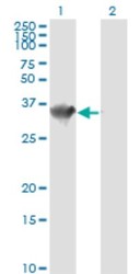
- Experimental details
- Western Blot: HADH Antibody (4B5) [H00003033-M01] - Analysis of HADH expression in transfected 293T cell line by HADHSC monoclonal antibody (M01), clone 4B5.Lane 1: HADH transfected lysate(34.3 KDa).Lane 2: Non-transfected lysate.
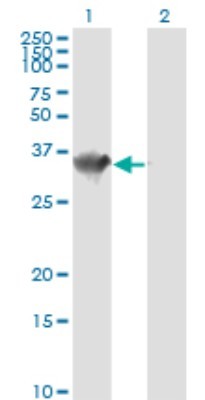
- Submitted by
- Novus Biologicals (provider)
- Main image
- Experimental details
- Western Blot: HADH Antibody (4B5) [H00003033-M01] - HADHSC monoclonal antibody (M01), clone 4B5 Analysis of HADHSC expression in HepG2.

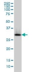
Supportive validation
- Submitted by
- Novus Biologicals (provider)
- Main image
- Experimental details
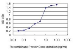
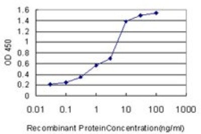
- ELISA: HADH Antibody (4B5) [H00003033-M01] - Detection limit for recombinant GST tagged HADHSC is approximately 0.03ng/ml as a capture antibody.
Supportive validation
- Submitted by
- Novus Biologicals (provider)
- Main image

- Experimental details
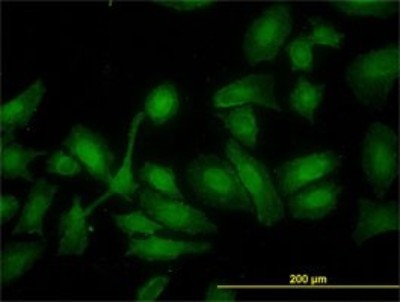
- Immunocytochemistry/Immunofluorescence: HADH Antibody (4B5) [H00003033-M01] - Analysis of monoclonal antibody to HADHSC on HeLa cell. Antibody concentration 10 ug/ml.
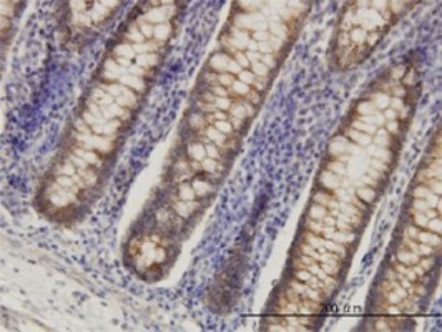
Supportive validation
- Submitted by
- Novus Biologicals (provider)
- Main image
- Experimental details
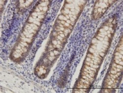
- Immunohistochemistry-Paraffin: HADH Antibody (4B5) [H00003033-M01] - Analysis of monoclonal antibody to HADHSC on formalin-fixed paraffin-embedded human colon. Antibody concentration 3 ug/ml.