 Explore
 Validate
 Learn
Learn Western blot
Antibody data
- Antibody Data
- Antigen structure
- References [4]
- Comments [0]
- Validations
- Western blot [3]
- Immunocytochemistry [2]
- Immunoprecipitation [2]
- Immunohistochemistry [1]
Submit
Validation data
Reference
Comment
Report error
- Product number
- GTX110568 - Provider product page

- Provider
- GeneTex
- Proper citation
- GeneTex Cat#GTX110568, RRID:AB_1949744
- Product name
- Bid antibody [N1C3-2]
- Antibody type
- Polyclonal
- Reactivity
- Human, Mouse, Rat
- Host
- Rabbit
Submitted references A high-throughput pipeline for validation of antibodies.
Dandelion root extract affects colorectal cancer proliferation and survival through the activation of multiple death signalling pathways.
Differentiation of human neuroblastoma cells toward the osteogenic lineage by mTOR inhibitor.
Generation of homologous cell pairs using the oral lymphatic system.
Sikorski K, Mehta A, Inngjerdingen M, Thakor F, Kling S, Kalina T, Nyman TA, Stensland ME, Zhou W, de Souza GA, Holden L, Stuchly J, Templin M, Lund-Johansen F
Nature methods 2018 Nov;15(11):909-912
Nature methods 2018 Nov;15(11):909-912
Dandelion root extract affects colorectal cancer proliferation and survival through the activation of multiple death signalling pathways.
Ovadje P, Ammar S, Guerrero JA, Arnason JT, Pandey S
Oncotarget 2016 Nov 8;7(45):73080-73100
Oncotarget 2016 Nov 8;7(45):73080-73100
Differentiation of human neuroblastoma cells toward the osteogenic lineage by mTOR inhibitor.
Carpentieri A, Cozzoli E, Scimeca M, Bonanno E, Sardanelli AM, Gambacurta A
Cell death & disease 2015 Nov 12;6(11):e1974
Cell death & disease 2015 Nov 12;6(11):e1974
Generation of homologous cell pairs using the oral lymphatic system.
Pu YF, Wang L, Wu HH, Bian H, Hong YY, Wang YX, Guo CB
International journal of clinical and experimental pathology 2014;7(4):1563-71
International journal of clinical and experimental pathology 2014;7(4):1563-71
No comments: Submit comment
Supportive validation
- Submitted by
- GeneTex (provider)
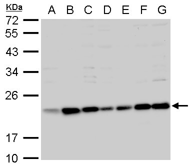
- Main image

- Experimental details
- Bid antibody [N1C3-2] detects Bid protein by western blot analysis.A. 30 ?g 293T whole cell lysate/extract B. 30 ?g A431 whole cell lysate/extract C. 30 ?g H1299 whole cell lysate/extract D. 30 ?g HeLa whole cell lysate/extract E. 30 ?g HepG2 whole cell lysate/extract F. 30 ?g Molt-4 whole cell lysate/extract G. 30 ?g Raji whole cell lysate/extract7.5 % SDS-PAGEBid antibody [N1C3-2] (GTX110568) dilution: 1:1000
- Submitted by
- GeneTex (provider)
- Main image
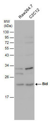
- Experimental details
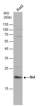
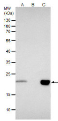
- Various whole cell extracts (30 £gg) were separated by 12% SDS-PAGE, and the membrane was blotted with Bid antibody [N1C3-2] (GTX110568) diluted at 1:500.
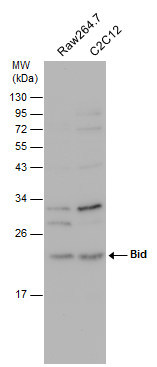
- Submitted by
- GeneTex (provider)
- Main image
- Experimental details
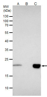
- Whole cell extract (30 £gg) was separated by 12% SDS-PAGE, and the membrane was blotted with Bid antibody [N1C3-2] (GTX110568) diluted at 1:1000.
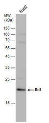
Supportive validation
- Submitted by
- GeneTex (provider)
- Main image
- Experimental details
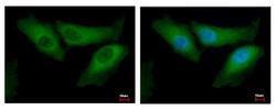
- Bid antibody [N1C3-2] detects BID protein at cytoplasm by immunofluorescent analysis. Sample: HeLa cells were fixed in 2% paraformaldehyde/culture medium at 37¢J for 30 min.Green: BID protein stained by Bid antibody [N1C3-2] (GTX110568) diluted at 1:500.Blue: Hoechst 33343 staining.
- Submitted by
- GeneTex (provider)
- Main image
- Experimental details
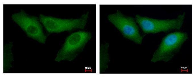
- Bid antibody [N1C3-2] detects Bid protein at cytoplasm by immunofluorescent analysis.Sample: A431 cells were fixed in 4% paraformaldehyde at RT for 15 min.Green: Bid protein stained by Bid antibody [N1C3-2] (GTX110568) diluted at 1:1000.Red: alpha Tubulin, a cytoskeleton marker, stained by alpha Tubulin antibody [GT114] (GTX628802) diluted at 1:1000.Blue: Hoechst 33342 staining.

Supportive validation
- Submitted by
- GeneTex (provider)
- Main image
- Experimental details
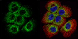
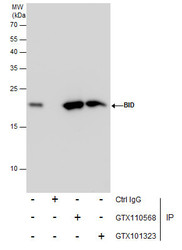
- Bid antibody [N1C3-2] immunoprecipitates BID protein in IP experiments.IP samples: Jurkat whole cell extractA. 40 ?g Jurkat whole cell extractB. Control with 4 ?g of preimmune Rabbit IgGC. Immunoprecipitation of BID protein by 4 ?g Bid antibody [N1C3-2] (GTX110568)5 % SDS-PAGEThe immunoprecipitated BID protein was detected by Bid antibody [N1C3-2] (GTX110568) diluted at 1:500.[EasyBlot anti-rabbit IgG (GTX221666-01) was used as a secondary reagent]
- Validation comment
- IP
- Submitted by
- GeneTex (provider)
- Main image
- Experimental details
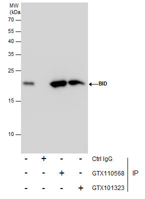
- Immunoprecipitation of Bid protein from Jurkat whole cell extracts using 5 £gg of Bid antibody [N1C3-2] (GTX110568) or Bid antibody [N1C3] (GTX101323).Western blot analysis was performed using Bid antibody [N1C3-2] (GTX110568).EasyBlot anti-Rabbit IgG (GTX221666-01) was used as a secondary reagent.
Supportive validation
- Submitted by
- GeneTex (provider)
- Main image
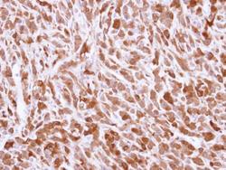
- Experimental details
- Immunohistochemical analysis of paraffin-embedded U87 xenograft, using Bid(GTX110568) antibody at 1:500 dilution.
