Explore
 Validate
 Learn
Learn Western blot
Antibody data
- Antibody Data
- Antigen structure
- References [7]
- Comments [0]
- Validations
- Western blot [1]
- Immunocytochemistry [1]
Submit
Validation data
Reference
Comment
Report error
- Product number
- 44-433G - Provider product page

- Provider
- Invitrogen Antibodies
- Product name
- BID p15 Polyclonal Antibody
- Antibody type
- Polyclonal
- Antigen
- Synthetic peptide
- Reactivity
- Human
- Host
- Rabbit
- Isotype
- IgG
- Vial size
- 100 µL
- Storage
- -20°C
Submitted references Caspase-4 is essential for saikosaponin a-induced apoptosis acting upstream of caspase-2 and γ-H2AX in colon cancer cells.
Sequential caspase-2 and caspase-8 activation is essential for saikosaponin a-induced apoptosis of human colon carcinoma cell lines.
Desferrioxamine (DFX) induces apoptosis through the p38-caspase8-Bid-Bax pathway in PHA-stimulated human lymphocytes.
Hypoxia/reoxygenation induces apoptosis through a ROS-mediated caspase-8/Bid/Bax pathway in human lymphocytes.
TRAIL/bortezomib cotreatment is potentially hepatotoxic but induces cancer-specific apoptosis within a therapeutic window.
Early growth response gene 1-mediated apoptosis is essential for transforming growth factor beta1-induced pulmonary fibrosis.
Adenoviral Bid overexpression induces caspase-dependent cleavage of truncated Bid and p53-independent apoptosis in human non-small cell lung cancers.
Kang SJ, Lee YJ, Kang SG, Cho S, Yoon W, Lim JH, Min SH, Lee TH, Kim BM
Oncotarget 2017 Nov 21;8(59):100433-100448
Oncotarget 2017 Nov 21;8(59):100433-100448
Sequential caspase-2 and caspase-8 activation is essential for saikosaponin a-induced apoptosis of human colon carcinoma cell lines.
Kim BM, Hong SH
Apoptosis : an international journal on programmed cell death 2011 Feb;16(2):184-97
Apoptosis : an international journal on programmed cell death 2011 Feb;16(2):184-97
Desferrioxamine (DFX) induces apoptosis through the p38-caspase8-Bid-Bax pathway in PHA-stimulated human lymphocytes.
Kim BM, Chung HW
Toxicology and applied pharmacology 2008 Apr 1;228(1):24-31
Toxicology and applied pharmacology 2008 Apr 1;228(1):24-31
Hypoxia/reoxygenation induces apoptosis through a ROS-mediated caspase-8/Bid/Bax pathway in human lymphocytes.
Kim BM, Chung HW
Biochemical and biophysical research communications 2007 Nov 23;363(3):745-50
Biochemical and biophysical research communications 2007 Nov 23;363(3):745-50
TRAIL/bortezomib cotreatment is potentially hepatotoxic but induces cancer-specific apoptosis within a therapeutic window.
Koschny R, Ganten TM, Sykora J, Haas TL, Sprick MR, Kolb A, Stremmel W, Walczak H
Hepatology (Baltimore, Md.) 2007 Mar;45(3):649-58
Hepatology (Baltimore, Md.) 2007 Mar;45(3):649-58
Early growth response gene 1-mediated apoptosis is essential for transforming growth factor beta1-induced pulmonary fibrosis.
Lee CG, Cho SJ, Kang MJ, Chapoval SP, Lee PJ, Noble PW, Yehualaeshet T, Lu B, Flavell RA, Milbrandt J, Homer RJ, Elias JA
The Journal of experimental medicine 2004 Aug 2;200(3):377-89
The Journal of experimental medicine 2004 Aug 2;200(3):377-89
Adenoviral Bid overexpression induces caspase-dependent cleavage of truncated Bid and p53-independent apoptosis in human non-small cell lung cancers.
Fukazawa T, Walter B, Owen-Schaub LB
The Journal of biological chemistry 2003 Jul 11;278(28):25428-34
The Journal of biological chemistry 2003 Jul 11;278(28):25428-34
No comments: Submit comment
Supportive validation
- Submitted by
- Invitrogen Antibodies (provider)
- Main image
- Experimental details
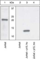
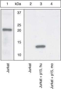
- Western Blot Extracts of Jurkat cells without added BID (lanes 1 and 2), with caspase-8 cleaved recombinant human BID (lane 3), or with caspase-8 cleaved recombinant mouse BID (lane 4) were resolved by SDS-PAGE on a 4-20% Tris-glycine gel and transferred to PVDF. The membrane was blocked with a 5% BSA-TBST buffer for one hour at room temperature, then incubated with either a human BID full length antibody (lane 1) or the human BID (p15) cleavage site-specific antibody (lanes 2-4) for two hours at room temperature in a 1% BSA-TBST buffer. After washing, the membrane was incubated with goat F (ab’)2 anti-rabbit IgG alkaline phosphatase (Product # ALI4405) and signals were detected using the Pierce SuperSignal™ method. The data show that the human BID (p15) antibody recognizes only the human 15 kDa BID fragment, demonstrating the specificity of the antibody.
Supportive validation
- Submitted by
- Invitrogen Antibodies (provider)
- Main image
- Experimental details
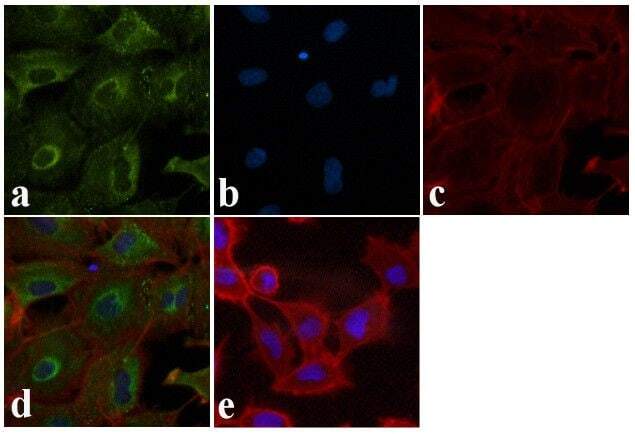
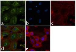
- Immunofluorescence analysis of BID (p15) was done on 70% confluent log phase A549 cells. The cells were fixed with 4% paraformaldehyde for 15 minutes, permeabilized with 0.25% Triton™ X-100 for 10 minutes, and blocked with 5% BSA for 1 hour at room temperature. The cells were labeled with BID (p15) Rabbit polyclonal Antibody (Product # 44-433G) at 1:250 dilution in 1% BSA and incubated for 3 hours at room temperature and then labeled with Alexa Fluor 488 Goat Anti-Rabbit IgG Secondary Antibody (Product # A-11008) at a dilution of 1:400 for 30 minutes at room temperature (Panel a: green). Nuclei (Panel b: blue) were stained with SlowFade® Gold Antifade Mountant DAPI (Product # S36938). F-actin (Panel c: red) was stained with Alexa Fluor 594 Phalloidin (Product # A12381). Panel d is a merged image showing cytoplasmic localization. Panel e shows no primary antibody control. The images were captured at 20X magnification.