Explore
 Validate
 Learn
Learn Western blot
Antibody data
- Antibody Data
- Antigen structure
- References [6]
- Comments [0]
- Validations
- Western blot [2]
Submit
Validation data
Reference
Comment
Report error
- Product number
- MAB860 - Provider product page

- Provider
- R&D Systems
- Product name
- Human/Mouse BID Antibody
- Antibody type
- Monoclonal
- Description
- Protein A or G purified from hybridoma culture supernatant. Detects human and mouse BID in Western blots. Detects full-length mouse BID and the 15 kDa carboxyl-terminal fragment (tBID) generated by cleavage with Caspase-8. The antibody also detects human BID. However, in Western blots of recombinant human and mouse BID, the antibody is most sensitive for mouse BID.
- Reactivity
- Human, Mouse
- Host
- Rat
- Conjugate
- Unconjugated
- Antigen sequence
P70444- Isotype
- IgG
- Antibody clone number
- 91508
- Vial size
- 100 ug
- Concentration
- LYOPH
- Storage
- Use a manual defrost freezer and avoid repeated freeze-thaw cycles. 12 months from date of receipt, -20 to -70 °C as supplied. 1 month, 2 to 8 °C under sterile conditions after reconstitution. 6 months, -20 to -70 °C under sterile conditions after reconstitution.
Submitted references Impact of caspase-1/11, -3, -7, or IL-1β/IL-18 deficiency on rabies virus-induced macrophage cell death and onset of disease.
Sensing cytosolic RpsL by macrophages induces lysosomal cell death and termination of bacterial infection.
The PI3K regulatory subunits p55α and p50α regulate cell death in vivo.
BH3-only protein Bid is dispensable for seizure-induced neuronal death and the associated nuclear accumulation of apoptosis-inducing factor.
Activation of the extrinsic caspase pathway in cultured cortical neurons requires p53-mediated down-regulation of the X-linked inhibitor of apoptosis protein to induce apoptosis.
Proteolysis of HIP during apoptosis occurs within a region similar to the BID loop.
Kip E, Nazé F, Suin V, Vanden Berghe T, Francart A, Lamoral S, Vandenabeele P, Beyaert R, Van Gucht S, Kalai M
Cell death discovery 2017;3:17012
Cell death discovery 2017;3:17012
Sensing cytosolic RpsL by macrophages induces lysosomal cell death and termination of bacterial infection.
Zhu W, Tao L, Quick ML, Joyce JA, Qu JM, Luo ZQ
PLoS pathogens 2015 Mar;11(3):e1004704
PLoS pathogens 2015 Mar;11(3):e1004704
The PI3K regulatory subunits p55α and p50α regulate cell death in vivo.
Pensa S, Neoh K, Resemann HK, Kreuzaler PA, Abell K, Clarke NJ, Reinheckel T, Kahn CR, Watson CJ
Cell death and differentiation 2014 Sep;21(9):1442-50
Cell death and differentiation 2014 Sep;21(9):1442-50
BH3-only protein Bid is dispensable for seizure-induced neuronal death and the associated nuclear accumulation of apoptosis-inducing factor.
Engel T, Caballero-Caballero A, Schindler CK, Plesnila N, Strasser A, Prehn JH, Henshall DC
Journal of neurochemistry 2010 Oct;115(1):92-101
Journal of neurochemistry 2010 Oct;115(1):92-101
Activation of the extrinsic caspase pathway in cultured cortical neurons requires p53-mediated down-regulation of the X-linked inhibitor of apoptosis protein to induce apoptosis.
Tun C, Guo W, Nguyen H, Yun B, Libby RT, Morrison RS, Garden GA
Journal of neurochemistry 2007 Aug;102(4):1206-19
Journal of neurochemistry 2007 Aug;102(4):1206-19
Proteolysis of HIP during apoptosis occurs within a region similar to the BID loop.
Caruso JA, Reiners JJ Jr
Apoptosis : an international journal on programmed cell death 2006 Nov;11(11):1877-85
Apoptosis : an international journal on programmed cell death 2006 Nov;11(11):1877-85
No comments: Submit comment
Supportive validation
- Submitted by
- R&D Systems (provider)
- Main image
- Experimental details
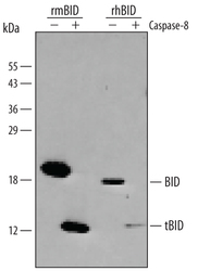
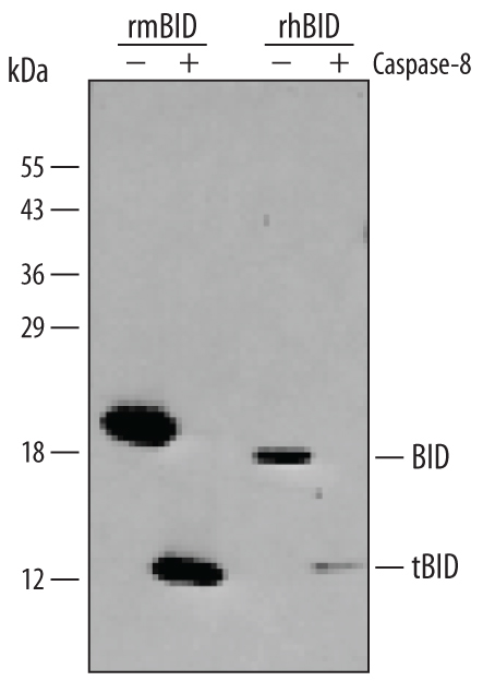
- Detection of Human and Mouse BID by Western Blot. Western blot shows lysates of Recombinant Mouse BID and Recombinant Human BID untreated (-) or treated (+) with Caspase-8. PVDF membrane was probed with 1 µg/mL Rat Anti-Human/Mouse BID Monoclonal Antibody (Catalog # MAB860) followed by HRP-conjugated Anti-Rat IgG Secondary Antibody (Catalog # HAF005). Specific bands for BID were detected at approximately 12 kDa and 20 kDa (as indicated). This experiment was conducted under reducing conditions and using Immunoblot Buffer Group 4.
- Submitted by
- R&D Systems (provider)
- Main image

- Experimental details
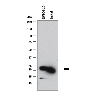
- Detection of Human and Mouse BID by Western Blot. Western blot shows lysates of SVEC4-10 mouse vascular endothelial cell line and Jurkat human acute T cell leukemia cell line. PVDF membrane was probed with 1 µg/mL of Rat Anti-Human/Mouse BID Monoclonal Antibody (Catalog # MAB860) followed by HRP-conjugated Anti-Rat IgG Secondary Antibody (Catalog # HAF005). A specific band was detected for BID at approximately 20 kDa (as indicated). This experiment was conducted under reducing conditions and using Immunoblot Buffer Group 1.