Explore
 Validate
 Learn
Learn Western blot
 Immunoprecipitation
Antibody data
- Antibody Data
- Antigen structure
- References [5]
- Comments [0]
- Validations
- Western blot [2]
- Immunohistochemistry [2]
Submit
Validation data
Reference
Comment
Report error
- Product number
- NB100-56106 - Provider product page

- Provider
- Novus Biologicals
- Proper citation
- Novus Cat#NB100-56106, RRID:AB_2065641
- Product name
- Rabbit Polyclonal BID Antibody
- Antibody type
- Polyclonal
- Description
- Unpurified. Full-length Bid is known to undergo cleavage/truncation (reviewed in Yin, 2006). Bid was initially found to be cleaved and activated by capspase-8 following death receptor activation. The term "Bid" was first used to described the caspase-8 cleaved/truncated C-terminal Bid. Bid can be also cleaved by other proteases such as Granzyme B, calpains and cathepsin. The C-terminal portion is considered to be the active Bid moiety. This active form can translocate from the cytosol to the mitochondria. Therefore, the appearance of Bid in the mitochondria is considered to be an indication of active Bid. However, it should also be noted that Bid has been shown to translocate to the mitochondria without cleavage in some model systems. The proteolytic cleavage of Bid usually occurs in the unstructured loop region between the alpha 2 and alpha 3 helices, which is between amino acids (aa) 41 and 79 of Bid. For example, the caspase-8/3 cleavage site is at 60 (human) and 59 (mouse). It should be noted that after cleavage, the smaller N-terminal portion of Bid is not necessarily separated from the larger C-terminal portion. The exact size of the C-terminal cleavage products depends on the Bid cleavage sites
- Reactivity
- Human, Mouse, Rat, Canine
- Host
- Rabbit
- Isotype
- IgG
- Vial size
- 0.05 ml
- Storage
- Store at 4C short term. Aliquot and store at -20C long term. Avoid freeze-thaw cycles.
Submitted references Effects of exosome-mediated delivery of myostatin propeptide on functional recovery of mdx mice.
Beclin-1-mediated Autophagy Protects Against Cadmium-activated Apoptosis via the Fas/FasL Pathway in Primary Rat Proximal Tubular Cell Culture.
Loss of α(E)-catenin promotes Fas mediated apoptosis in tubular epithelial cells.
Dexamethasone decreases cholestatic liver injury via inhibition of intrinsic pathway with simultaneous enhancement of mitochondrial biogenesis.
Early processing of Bid and caspase-6, -8, -10, -14 in the canine brain during cardiac arrest and resuscitation.
Ran N, Gao X, Dong X, Li J, Lin C, Geng M, Yin H
Biomaterials 2020 Apr;236:119826
Biomaterials 2020 Apr;236:119826
Beclin-1-mediated Autophagy Protects Against Cadmium-activated Apoptosis via the Fas/FasL Pathway in Primary Rat Proximal Tubular Cell Culture.
Liu G, Yuan Y, Long M, Luo T, Bian J, Liu X, Gu J, Zou H, Song R, Wang Y, Wang L, Liu Z
Scientific reports 2017 Apr 20;7(1):977
Scientific reports 2017 Apr 20;7(1):977
Loss of α(E)-catenin promotes Fas mediated apoptosis in tubular epithelial cells.
Wang X, Parrish AR
Apoptosis : an international journal on programmed cell death 2015 Jul;20(7):921-9
Apoptosis : an international journal on programmed cell death 2015 Jul;20(7):921-9
Dexamethasone decreases cholestatic liver injury via inhibition of intrinsic pathway with simultaneous enhancement of mitochondrial biogenesis.
Tiao MM, Lin TK, Chen JB, Liou CW, Wang PW, Huang CC, Chou YM, Huang YH, Chuang JH
Steroids 2011 Jun;76(7):660-6
Steroids 2011 Jun;76(7):660-6
Early processing of Bid and caspase-6, -8, -10, -14 in the canine brain during cardiac arrest and resuscitation.
Krajewska M, Rosenthal RE, Mikolajczyk J, Stennicke HR, Wiesenthal T, Mai J, Naito M, Salvesen GS, Reed JC, Fiskum G, Krajewski S
Experimental neurology 2004 Oct;189(2):261-79
Experimental neurology 2004 Oct;189(2):261-79
No comments: Submit comment
Supportive validation
- Submitted by
- Novus Biologicals (provider)
- Main image

- Experimental details
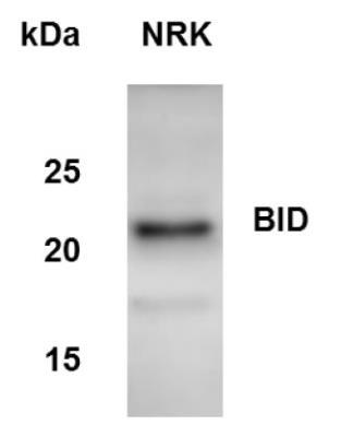
- Western Blot: BID Antibody [NB100-56106] - Analysis of BID in NRK whole cell lysate using anti-BID antibody. Image from verified customer review.
- Submitted by
- Novus Biologicals (provider)
- Main image
- Experimental details
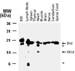
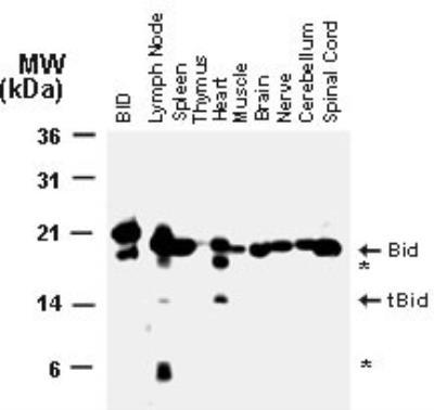
- Western Blot: BID Antibody [NB100-56106] - Analysis of Bid in normal mouse tissues using this antibody. BID = recombinant Bid. Arrowheads indicate the positions of the full-length (uncleaved) ~22 kDa Bid and the ~15 kDa truncated form of Bid (tBid) typical of the caspase-cleavage. Additional bands representing partial Bid degradation products are indicated by asterisks (*).
Supportive validation
- Submitted by
- Novus Biologicals (provider)
- Main image
- Experimental details
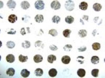
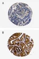
- Immunohistochemistry-Paraffin: BID Antibody [NB100-56106] - Two cores from a human ovarian carcinoma formalin-fixed, paraffin-embedded tissue microarray demonstrate the variable expression of Bid protein. The sections were stained with Bid antibody at 1:2000 with hematoxylin-eosin counterstain. Section A shows very weak staining while section B stains much more strongly.

- Submitted by
- Novus Biologicals (provider)
- Main image

- Experimental details
- Immunohistochemistry-Paraffin: BID Antibody [NB100-56106] - Formalin-fixed, paraffin-embedded human ovarian carcinoma tissue array stained for Bid expression using this antibody at 1:2000. Hematoxylin-eosin counterstain. Variable Bid expression is seen between patient samples.