Explore
 Validate
 Learn
Learn Western blot
 ELISA
ELISAAntibody data
- Antibody Data
- Antigen structure
- References [0]
- Comments [0]
- Validations
- Western blot [1]
- Immunohistochemistry [2]
Submit
Validation data
Reference
Comment
Report error
- Product number
- NBP1-78017 - Provider product page

- Provider
- Novus Biologicals
- Proper citation
- Novus Cat#NBP1-78017, RRID:AB_11027576
- Product name
- Rabbit Polyclonal PERK Antibody
- Antibody type
- Polyclonal
- Description
- Unpurified.
- Reactivity
- Mouse
- Host
- Rabbit
- Vial size
- 0.1 ml
- Storage
- Store at -20C. Avoid freeze-thaw cycles.
No comments: Submit comment
Supportive validation
- Submitted by
- Novus Biologicals (provider)
- Main image

- Experimental details
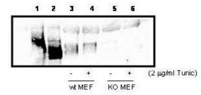
- Western Blot: PERK Antibody [NBP1-78017] - PERK in cell lysates. 300ug PERK over-expressing 293T cell lysate (lanes 1 & 2), or 800ug wild type (Lanes 3 & 4), and PERK knock out (lanes 5 & 6) MEF cell lysate were immunoprecipated with 15ul anti-PERK, followed by western blotIgG with 1:1000 dilution of anti-PERK in 5% milk/TBST buffer. Lane 1, 293T cells over-expressing Myc-PERK wt, Lane 2, 293T cells over-expressing Myc-PERK K618A. Personal Communication. A, Diehl, Univ. of Pennsylvania, Philadelphia, PA.
Supportive validation
- Submitted by
- Novus Biologicals (provider)
- Main image
- Experimental details
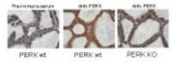
- Immunohistochemistry-Paraffin: PERK Antibody [NBP1-78017] - Immunohistochemistry staining of mouse mammary gland samples from lactating mice (L10) with anti-PERK. Positive staining signal observed in wild type mouse sample with anti-PERK staining only (middle image), but not in the knock out mouse sample (right image) and pre-immune serum staining (left image) The anti-PERK was diluted 1:1,000 in 5% goat serum in PBS and allowed to incubate for 2h at room temperature in a humidified chamber. Personal Communication. A, Diehl, Univ. of Pennsylvania, Philadelphia, PA.
- Submitted by
- Novus Biologicals (provider)
- Main image

- Experimental details
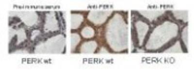
- Immunohistochemistry: PERK Antibody [NBP1-78017] - Analysis of mouse mammary gland samples from lactating mice (L10) with anti-PERK. Positive staining signal observed in wild type mouse sample with anti-PERK staining only (middle image), but not in the knock out mouse sample (right image) and pre-immune serum staining (left image) The anti-PERK was diluted 1:1,000 in 5% goat serum in PBS and allowed to incubate for 2h at room temperature in a humidified chamber