Explore
 Validate
 Learn
Learn Western blot
 Immunocytochemistry
 Immunoprecipitation
Antibody data
- Antibody Data
- Antigen structure
- References [0]
- Comments [0]
- Validations
- Western blot [1]
- Immunoprecipitation [3]
- Immunohistochemistry [2]
Submit
Validation data
Reference
Comment
Report error
- Product number
- LS-C353177 - Provider product page

- Provider
- LSBio
- Product name
- EIF2AK3 / PERK Antibody (N-Terminus) LS-C353177
- Antibody type
- Polyclonal
- Description
- Immunoaffinity purified
- Reactivity
- Human
- Host
- Rabbit
- Storage
- Store at -20°C. Aliquot to avoid freeze/thaw cycles.
No comments: Submit comment
Enhanced validation
- Submitted by
- LSBio (provider)
- Enhanced method
- Genetic validation
- Main image

- Experimental details
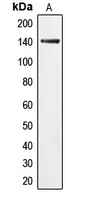
- Western blot analysis of PERK expression in HeLa (A) whole cell lysates.
Supportive validation
- Submitted by
- LSBio (provider)
- Enhanced method
- Genetic validation
- Main image

- Experimental details
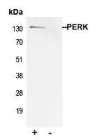
- Immunoprecipitation of PERK from 0.5mg HeLa whole cell extract lysate using 5ug of Anti-PERK Antibody and 50ul of protein G magnetic beads (+). No antibody was added to the control (-). The antibody was incubated under agitation with Protein G beads for 10min HeLa whole cell extract lysate diluted in RIPA buffer was added to each sample and incubated for a further 10min under agitation. Proteins were eluted by addition of 40ul SDS loading buffer and incubated for 10min at 70 C; 10ul of each sample was separated on a SDS PAGE gel transferred to a nitrocellulose membrane blocked with 5% BSA and probed with Anti-PERK Antibody.
- Submitted by
- LSBio (provider)
- Main image

- Experimental details
- Immunoprecipitation of PERK from 0.5mg HeLa whole cell extract lysate using 5ug of Anti-PERK Antibody and 50ul of protein G magnetic beads (+). No antibody was added to the control (-). The antibody was incubated under agitation with Protein G beads for 10min HeLa whole cell extract lysate diluted in RIPA buffer was added to each sample and incubated for a further 10min under agitation. Proteins were eluted by addition of 40ul SDS loading buffer and incubated for 10min at 70 C; 10ul of each sample was separated on a SDS PAGE gel transferred to a nitrocellulose membrane blocked with 5% BSA and probed with Anti-PERK Antibody.

Supportive validation
- Submitted by
- LSBio (provider)
- Enhanced method
- Genetic validation
- Main image

- Experimental details
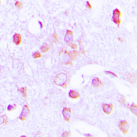
- Immunohistochemical analysis of PERK staining in human brain formalin fixed paraffin embedded tissue section. The section was pre-treated using heat mediated antigen retrieval with sodium citrate buffer (pH 6.0). The section was then incubated with the antibody at room temperature and detected using an HRP conjugated compact polymer system. DAB was used as the chromogen. The section was then counterstained with hematoxylin and mounted with DPX.

- Submitted by
- LSBio (provider)
- Enhanced method
- Genetic validation
- Main image
- Experimental details
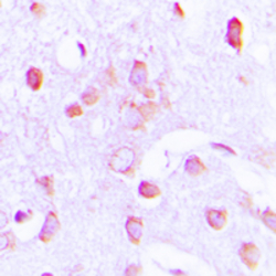
- Immunohistochemical analysis of PERK staining in human brain formalin fixed paraffin embedded tissue section. The section was pre-treated using heat mediated antigen retrieval with sodium citrate buffer (pH 6.0). The section was then incubated with the antibody at room temperature and detected using an HRP conjugated compact polymer system. DAB was used as the chromogen. The section was then counterstained with hematoxylin and mounted with DPX.