Explore
 Validate
 Learn
Learn Western blot
 ELISA
ELISAAntibody data
- Antibody Data
- Antigen structure
- References [6]
- Comments [0]
- Validations
- Western blot [1]
Submit
Validation data
Reference
Comment
Report error
- Product number
- A01992-2 - Provider product page

- Provider
- Boster Biological Technology
- Product name
- Anti-PERK/EIF2AK3 Antibody Picoband™
- Antibody type
- Polyclonal
- Description
- Polyclonal antibody for PERK/EIF2AK3 detection. Host: Rabbit.Size: 100μg/vial. Tested applications: WB, FCM, Direct ELISA. Reactive species: Human. PERK/EIF2AK3 information: Subcellular Localization: Endoplasmic reticulum membrane; Single-pass type I membrane protein; Tissue Specificity: Ubiquitous. A high level expression is seen in secretory tissues.
- Reactivity
- Human, Mouse, Rat
- Host
- Rabbit
- Vial size
- 100μg/vial
- Concentration
- 0.5-1mg/ml, actual concentration vary by lot. Use suggested dilution ratio to decide dilution procedure.
- Storage
- At -20°C for one year. After reconstitution, at 4°C for one month. It can also be aliquoted and stored frozen at -20°C for a longer time. Avoid repeated freezing and thawing.
- Handling
- Add 0.2ml of distilled water will yield a concentration of 500ug/ml.
Submitted references Total Panax notoginseng saponin inhibits balloon injury-induced neointimal hyperplasia in rat carotid artery models by suppressing pERK/p38 MAPK pathways.
ASK1 Enhances Angiotensin II-Induced Liver Fibrosis In Vitro by Mediating Endoplasmic Reticulum Stress-Dependent Exosomes.
miR-9-5p attenuates ischemic stroke through targeting ERMP1-mediated endoplasmic reticulum stress.
Effects of 5-hydroxy-4'-nitro-7-propionyloxy-genistein on inhibiting proliferation and invasion via activating reactive oxygen species in human ovarian cancer A2780/DDP cells.
Recombinant Newcastle disease virus (rL-RVG) triggers autophagy and apoptosis in gastric carcinoma cells by inducing ER stress.
Endoplasmic reticulum stress in diethylnitrosamine-induced rat liver cancer.
Yang Z, Zhang H, An M, Bian M, Song M, Guo X, Liu Q, Qiu M
Brazilian journal of medical and biological research = Revista brasileira de pesquisas medicas e biologicas 2020;53(1):e9085
Brazilian journal of medical and biological research = Revista brasileira de pesquisas medicas e biologicas 2020;53(1):e9085
ASK1 Enhances Angiotensin II-Induced Liver Fibrosis In Vitro by Mediating Endoplasmic Reticulum Stress-Dependent Exosomes.
Fang PP, Pan CW, Lin W, Li J, Huang SS, Zhou GY, Du WJ, Li Q
Mediators of inflammation 2020;2020:8183713
Mediators of inflammation 2020;2020:8183713
miR-9-5p attenuates ischemic stroke through targeting ERMP1-mediated endoplasmic reticulum stress.
Chi L, Jiao D, Nan G, Yuan H, Shen J, Gao Y
Acta histochemica 2019 Nov;121(8):151438
Acta histochemica 2019 Nov;121(8):151438
Effects of 5-hydroxy-4'-nitro-7-propionyloxy-genistein on inhibiting proliferation and invasion via activating reactive oxygen species in human ovarian cancer A2780/DDP cells.
Bai J, Yang BJ, Luo X
Oncology letters 2018 Apr;15(4):5227-5235
Oncology letters 2018 Apr;15(4):5227-5235
Recombinant Newcastle disease virus (rL-RVG) triggers autophagy and apoptosis in gastric carcinoma cells by inducing ER stress.
Bu X, Zhao Y, Zhang Z, Wang M, Li M, Yan Y
American journal of cancer research 2016;6(5):924-36
American journal of cancer research 2016;6(5):924-36
Endoplasmic reticulum stress in diethylnitrosamine-induced rat liver cancer.
Xiao B, Cui LM, Ma DJ, Liu SP, Zhang XW
Oncology letters 2014 Jan;7(1):23-27
Oncology letters 2014 Jan;7(1):23-27
No comments: Submit comment
Supportive validation
- Submitted by
- Boster Biological Technology (provider)
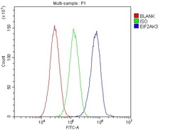
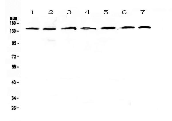
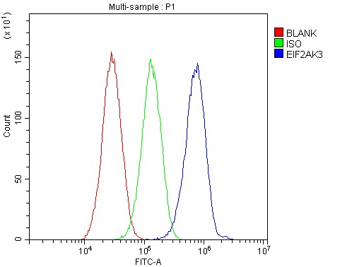
- Main image
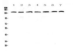
- Experimental details
- Western blot analysis of PERK using anti-PERK antibody (A01992-2). Electrophoresis was performed on a 5-20% SDS-PAGE gel at 70V (Stacking gel) / 90V (Resolving gel) for 2-3 hours. The sample well of each lane was loaded with 50ug of sample under reducing conditions. Lane 1: human Hela whole cell lysates,Lane 2: human COLO-320 whole cell lysates,Lane 3: human A549 whole cell lysates,Lane 4: human SK-OV-3 whole cell lysates,Lane 5: Human A431 whole cell lysates,Lane 6: rat brain tissue lysates,Lane 7: mouse brain tissue lysates. After Electrophoresis, proteins were transferred to a Nitrocellulose membrane at 150mA for 50-90 minutes. Blocked the membrane with 5% Non-fat Milk/ TBS for 1.5 hour at RT. The membrane was incubated with rabbit anti-PERK antigen affinity purified polyclonal antibody (Catalog # A01992-2) at 0.5 μg/mL overnight at 4°C, then washed with TBS-0.1%Tween 3 times with 5 minutes each and probed with a goat anti-rabbit IgG-HRP secondary antibody at a dilution of 1:10000 for 1.5 hour at RT. The signal is developed using an Enhanced Chemiluminescent detection (ECL) kit (Catalog # EK1002) with Tanon 5200 system. A specific band was detected for PERK at approximately 140KD. The expected band size for PERK is at 125KD.
- Additional image