Explore
 Validate
 Learn
Learn Western blot
 Immunohistochemistry
Antibody data
- Antibody Data
- Antigen structure
- References [1]
- Comments [0]
- Validations
- Immunohistochemistry [1]
- Other assay [3]
Submit
Validation data
Reference
Comment
Report error
- Product number
- PA5-34461 - Provider product page

- Provider
- Invitrogen Antibodies
- Product name
- CMPK2 Polyclonal Antibody
- Antibody type
- Polyclonal
- Antigen
- Synthetic peptide
- Description
- A suggested positive control is rat lung tissue lysate. PA5-34461 can be used with blocking peptide PEP-1503.
- Reactivity
- Human, Mouse, Rat
- Host
- Rabbit
- Isotype
- IgG
- Vial size
- 100 μg
- Concentration
- 1 mg/mL
- Storage
- Maintain refrigerated at 2-8°C for up to 3 months. For long term storage store at -20°C
Submitted references Electroacupuncture Inhibits NLRP3 Activation by Regulating CMPK2 After Spinal Cord Injury.
Chen Y, Wu L, Shi M, Zeng D, Hu R, Wu X, Han S, He K, Xu H, Shao X, Ma R
Frontiers in immunology 2022;13:788556
Frontiers in immunology 2022;13:788556
No comments: Submit comment
Supportive validation
- Submitted by
- Invitrogen Antibodies (provider)
- Main image
- Experimental details
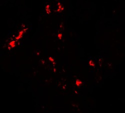
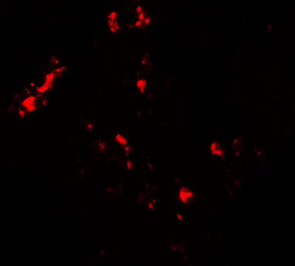
- Immunofluorescence of CMPK2 in human lung tissue with CMPK2 Polyclonal Antibody (Product # PA5-34461) at 20 µg/mL.
Supportive validation
- Submitted by
- Invitrogen Antibodies (provider)
- Main image
- Experimental details
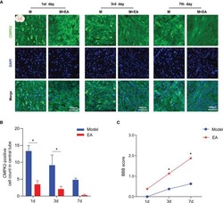
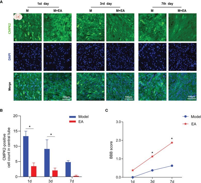
- Immunofluorescence analysis showed positive expression of CMPK2 in spinal cord tissues of the model and EA groups. (A) Immunofluorescence labeling represented the number of CMPK2-positive cells (green fluorescence) at 1, 3, and 7 days after SCI, and the CMPK2-positive protein staining area is shown in green. (B) Quantitative analysis of CMPK2-positive cells. *p < 0.05 compared with the model group; one-way anOVA was used for comparison. N = 5 rats/group. (C) BBB scores for the model and EA groups. N = 8 rats/group. * p < 0.05 compared with the sham group. Data are presented as the mean +- SEM. EA, electroacupuncture; BBB, Basso-Beattie-Bresnahan.
- Submitted by
- Invitrogen Antibodies (provider)
- Main image
- Experimental details
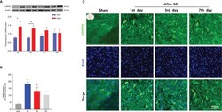
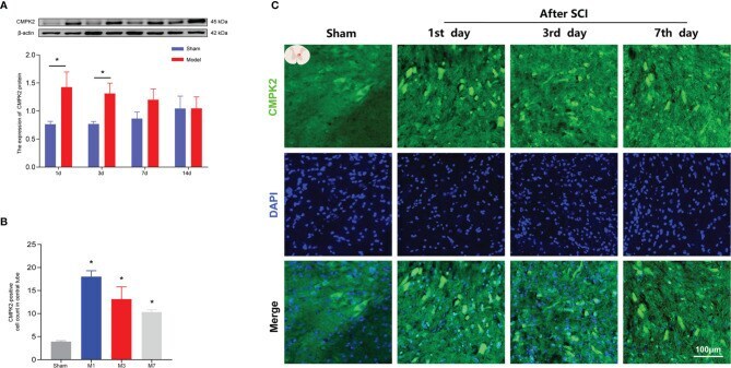
- The expression of CMPK2 in rat spinal cords was detected using WB and IF. (A) Western blotting was used to analyze the protein expression of CMPK2 in the sham and model groups after SCI. Representative protein bands are shown for CMPK at 1, 3, 7, and 14 days after SCI (N = 5 rats/group). *p < 0.05; two-way ANOVA was used for comparison. (B) Quantitative analysis of CMPK2-positive cells. *p < 0.05 compared with the sham group; one-way anOVA was used for comparison. N = 5 rats/group. (C) CMPK2 was expressed around the central canal of the gray matter of the spinal cord at 1, 3, and 7 days after spinal cord injury. CMPK2-positive cells are represented by green fluorescence. DAPI (blue) co-staining was used to identify positive cells. Data are presented as the mean +- SEM. WB, Western blotting; IF, immunofluorescence; SCI, spinal cord injury.
- Submitted by
- Invitrogen Antibodies (provider)
- Main image
- Experimental details
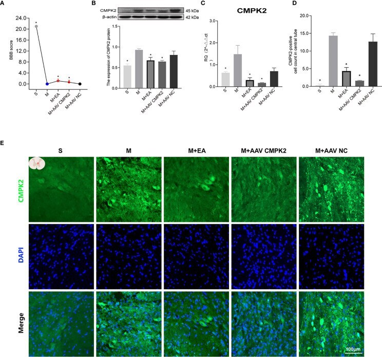
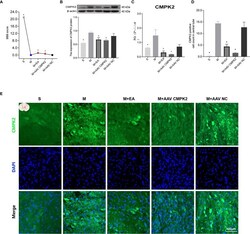
- Effect of EA on CMPK2 expression in spinal cords of rats following CMPK2 knockdown. (A) BBB scores of rats in the Sham, Model, M+EA, M+AAV CMPK2, and M+AAV NC groups. N = 8. *p < 0.05 compared with the model group. (B) Western blotting analysis of CMPK2 expression in spinal cord tissues of rats in the Sham, Model, M+EA, M+AAV CMPK2, and M+AAV NC groups. A representative Western blotting and semiquantitative analysis of CMPK2 are shown. N = 5 rats/group. * p < 0.05 compared with the model group; one-way anOVA was used for comparison. (C) Evaluation of CMPK2 gene expression using qPCR in the Sham, Model, M+EA, M+AAV CMPK2, and M+AAV NC groups 3 days after SCI. N = 5 rats/group. * p < 0.05 compared with the model group; one-way anOVA was used for comparison. (D) Quantitative analysis of CMPK2-positive cells. * p < 0.05 compared with the model group; one-way anOVA was used for comparison. N = 5 rats/group. (E) Immunofluorescence was used to detect the expression of CMPK2 around the central canal of the spinal gray matter of rats in the Sham, Model, M+EA, M+AAV CMPK2, and M+AAV NC groups. Number of CMPK2-positive cells (green fluorescence) 3 days after SCI. DAPI (blue) co-staining was used to identify positive cells. All data are expressed as the mean +- SEM. EA, electroacupuncture; BBB, Basso-Beattie-Bresnahan; SCI, spinal cord injury.