 Explore
 Validate
 Learn
Learn Western blot
 Immunocytochemistry
Antibody data
- Antibody Data
- Antigen structure
- References [1]
- Comments [0]
- Validations
- Immunocytochemistry [1]
- Immunohistochemistry [1]
Submit
Validation data
Reference
Comment
Report error
- Product number
- HPA054104 - Provider product page

- Provider
- Atlas Antibodies
- Proper citation
- Atlas Antibodies Cat#HPA054104, RRID:AB_2682380
- Product name
- Anti-EPB41L1
- Antibody type
- Polyclonal
- Description
- Polyclonal Antibody against Human EPB41L1, Gene description: erythrocyte membrane protein band 4.1-like 1, Alternative Gene Names: KIAA0338, Validated applications: WB, IHC, ICC, Uniprot ID: Q9H4G0, Storage: Store at +4°C for short term storage. Long time storage is recommended at -20°C.
- Reactivity
- Human
- Host
- Rabbit
- Conjugate
- Unconjugated
- Isotype
- IgG
- Vial size
- 100 µl
- Concentration
- 0.2 mg/ml
- Storage
- Store at +4°C for short term storage. Long time storage is recommended at -20°C.
- Handling
- The antibody solution should be gently mixed before use.
Submitted references Abnormal expression and prognostic significance of EPB41L1 in kidney renal clear cell carcinoma based on data mining
Liang T, Sang S, Shao Q, Chen C, Deng Z, Wang T, Kang Q
Cancer Cell International 2020;20(1)
Cancer Cell International 2020;20(1)
No comments: Submit comment
Supportive validation
- Submitted by
- Atlas Antibodies (provider)
- Main image
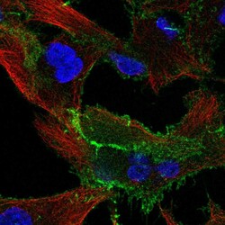
- Experimental details
- Immunofluorescent staining of human cell line A549 shows localization to plasma membrane.
- Sample type
- Human

Supportive validation
- Submitted by
- Atlas Antibodies (provider)
- Enhanced method
- Orthogonal validation
- Main image
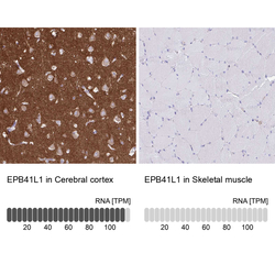
- Experimental details
- Immunohistochemistry analysis in human cerebral cortex and skeletal muscle tissues using HPA054104 antibody. Corresponding EPB41L1 RNA-seq data are presented for the same tissues.
- Sample type
- Human
- Protocol
- Protocol
