 Explore
 Validate
 Learn
LearnPA5-77728
antibody from Invitrogen Antibodies
Targeting: SCN8A
CerIII, CIAT, MED, NaCh6, Nav1.6, PN4
 Western blot
Antibody data
- Antibody Data
- Antigen structure
- References [0]
- Comments [0]
- Validations
- Western blot [1]
- Immunocytochemistry [3]
- Immunohistochemistry [1]
Submit
Validation data
Reference
Comment
Report error
- Product number
- PA5-77728 - Provider product page

- Provider
- Invitrogen Antibodies
- Product name
- SCN8A Polyclonal Antibody
- Antibody type
- Polyclonal
- Antigen
- Synthetic peptide
- Description
- For reconstitution, we recommend adding 100 µL distilled water to a final antibody concentration of about 1 mg/mL. To use this carrier-free antibody for conjugation experiments, we strongly recommend performing another round of desalting. (Zeba Spin Desalting Columns, 7KMWCO, 0.5 mL, Product # 89882)
- Reactivity
- Human, Mouse, Rat
- Host
- Rabbit
- Isotype
- IgG
- Vial size
- 50 µL
- Concentration
- 0.8 mg/mL
- Storage
- -20°C
No comments: Submit comment
Supportive validation
- Submitted by
- Invitrogen Antibodies (provider)
- Main image

- Experimental details
- Western blot analysis of rat brain membrane with SCN8A polyclonal antibody (Product # PA5-77728) using a dilution of 1:200.

Supportive validation
- Submitted by
- Invitrogen Antibodies (provider)
- Main image
- Experimental details
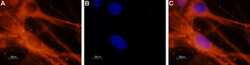
- Immunocytochemistry analysis of SCN8A in paraformaldehyde-fixed and permeabilized rat dorsal root ganglion cells. A) Samples were probed with SCN8A polyclonal antibody (Product # PA5-77728) at a dilution of 1:200, and incubated with goat-anti-rabbit-AlexaFluor-555 and Hoechst. B) Nuclei stained image. C) Merged images of Panels A and B.

- Submitted by
- Invitrogen Antibodies (provider)
- Main image
- Experimental details
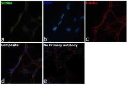
- Immunofluorescence analysis of SCN8A was performed using 70% confluent log phase IMR-32 cells. The cells were fixed with 4% paraformaldehyde for 10 minutes, permeabilized with 0.1% Triton™ X-100 for 15 minutes, and blocked with 2% BSA for 1 hour at room temperature. The cells were labeled with SCN8A Polyclonal Antibody (Product # PA5-77728) at 1:100 dilution in 0.1% BSA, incubated at 4 degree celsius overnight and then with Goat anti-Rabbit IgG (H+L), Superclonal™ Recombinant Secondary Antibody, Alexa Fluor 488 conjugate (Product # A27034) at a dilution of 1:2000 for 45 minutes at room temperature (Panel a: green). Nuclei (Panel b: blue) were stained with SlowFade® Gold Antifade Mountant with DAPI (Product # S36938). F-actin (Panel c: red) was stained with Rhodamine Phalloidin (Product # R415, 1:300). Panel d represents the merged image showing staining in plasma membrane. Panel e represents control cells with no primary antibody to assess the background. The images were captured at 60X magnification.

- Submitted by
- Invitrogen Antibodies (provider)
- Main image
- Experimental details
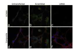
- Knockdown of SCN8A was achieved by transfecting IMR-32 cells with SCN8A specific siRNA (Silencer® select Product # s12535, s12534). Immunofluorescence analysis was performed on IMR-32 cells (untransfected, panel a,d), transfected with non-specific scrambled siRNA (panels b,e) and transfected with SCN8A specific siRNA (panel c,f). Cells were fixed, permeabilized, and labelled with SCN8A Polyclonal Antibody (Product # PA5-77728, 1:100 dilution), followed by Goat anti-Rabbit IgG (H+L) Superclonal™ Recombinant Secondary Antibody, Alexa Fluor® 488 conjugate (Product # A27034, 1:2000). Nuclei (blue) were stained using ProLong™ Diamond Antifade Mountant with DAPI (Product # P36962), and Rhodamine Phalloidin (Product # R415, 1:300) was used for cytoskeletal F-actin (red) staining. Reduction of specific signal was observed upon siRNA mediated knockdown (panel c,f) confirming specificity of the antibody to SCN8A (green). The images were captured at 60X magnification.

Supportive validation
- Submitted by
- Invitrogen Antibodies (provider)
- Main image

- Experimental details
- Immunohistochemistry analysis of SCN8A in mouse hippocampus. A) Samples were probed with SCN8A polyclonal antibody (Product # PA5-77728) using a dilution of 1:100, and incubated with parvalbumin and DAPI (green). B) Interneurons stained image (red). C) Merged image of Panels A and B.
