 Explore
 Validate
 Learn
LearnPA5-47441
antibody from Invitrogen Antibodies
Targeting: NECTIN3
CD113, CDw113, DKFZP566B0846, nectin-3, PPR3, PVRL3, PVRR3
 Western blot
 Immunocytochemistry
Antibody data
- Antibody Data
- Antigen structure
- References [0]
- Comments [0]
- Validations
- Immunocytochemistry [2]
- Immunohistochemistry [1]
Submit
Validation data
Reference
Comment
Report error
- Product number
- PA5-47441 - Provider product page

- Provider
- Invitrogen Antibodies
- Product name
- Nectin 3 Polyclonal Antibody
- Antibody type
- Polyclonal
- Antigen
- Recombinant full-length protein
- Description
- In direct ELISAs, less than 65% cross-reactivity with recombinant mouse Nectin-3 is observed and less than 5% cross-reactivity with recombinant human (rh) Nectin-1, rhNectin-2, and rhNectin-4 is observed. Reconstitute at 0.2 mg/mL in sterile PBS.
- Reactivity
- Human
- Host
- Goat
- Isotype
- IgG
- Vial size
- 100 µg
- Concentration
- 0.2 mg/mL
- Storage
- -20° C, Avoid Freeze/Thaw Cycles
No comments: Submit comment
Supportive validation
- Submitted by
- Invitrogen Antibodies (provider)
- Main image

- Experimental details
- Immunofluorescence analysis of Nectin 3 was performed using 70% confluent log phase Caco-2 cells. The cells were fixed with 4% paraformaldehyde for 10 minutes, permeabilized with 0.1% Triton™ X-100 for 15 minutes, and blocked with 2% BSA for 1 hour at room temperature. The cells were labeled with Nectin 3 Goat Polyclonal Antibody (Product # PA5-47441) at 5 microgram/mL in 0.1% BSA, incubated at 4 degree Celsius overnight and then labeled with Rabbit anti-Goat IgG (H+L) Cross-Adsorbed Secondary Antibody, Alexa Fluor 488 (Product # A-11078) at a dilution of 1:2000 for 45 minutes at room temperature (Panel a: green). Nuclei (Panel b: blue) were stained with ProLong™ Diamond Antifade Mountant with DAPI (Product # P36962). F-actin (Panel c: red) was stained with Rhodamine Phalloidin (Product # R415). Panel d represents the merged image showing membrane and cell junctional localization. Panel e represents control cells with no primary antibody to assess background. The images were captured at 60X magnification.

- Submitted by
- Invitrogen Antibodies (provider)
- Main image
- Experimental details
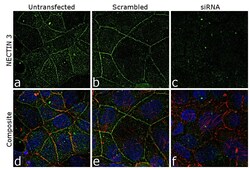
- KD of Nectin 3 was achieved by transfecting Caco-2 cells with Nectin 3 specific siRNA (Silencer® select Product # S24804). Immunofluorescence analysis was performed on untransfected Caco-2 cells (panel a,d), transfected with non-specific scrambled siRNA (panels b,e) and transfected with Nectin 3 specific siRNA (panel c,f). Cells were fixed, permeabilized, and labelled with Nectin 3 Goat Polyclonal Antibody (Product # PA5-47441, 5 µg/mL), followed by Rabbit anti-Goat IgG (H+L) Cross-Adsorbed Secondary Antibody, Alexa Fluor 488 (Product # A-11078, 1:2000). Nuclei (blue) were stained using ProLong™ Diamond Antifade Mountant with DAPI (Product # P36962), and Rhodamine Phalloidin (Product # R415, 1:300) was used for cytoskeletal F-actin (red) staining. Reduction of specific signal was observed upon siRNA mediated KD (panel c,f) confirming specificity of the antibody to Nectin 3 (green). The images were captured at 60X magnification.

Supportive validation
- Submitted by
- Invitrogen Antibodies (provider)
- Main image
- Experimental details
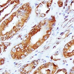
- Immunohistochemical analysis of Nectin 3 in immersion fixed paraffin-embedded sections of human kidney array. Samples were incubated in Nectin 3 polyclonal antibody (Product # PA5-47441) using a dilution of 10 µg/mL overnight at 4 °C. Tissue was stained using the Anti-Goat HRP-DAB Cell & Tissue Staining Kit (brown) and counterstained with hematoxylin (blue).
