Explore
 Validate
 Learn
Learn Western blot
 Immunocytochemistry
Antibody data
- Antibody Data
- Antigen structure
- References [1]
- Comments [0]
- Validations
- Immunocytochemistry [1]
- Immunohistochemistry [1]
Submit
Validation data
Reference
Comment
Report error
- Product number
- HPA027395 - Provider product page

- Provider
- Atlas Antibodies
- Proper citation
- Atlas Antibodies Cat#HPA027395, RRID:AB_10601554
- Product name
- Anti-OXR1
- Antibody type
- Polyclonal
- Description
- Polyclonal Antibody against Human OXR1, Gene description: oxidation resistance 1, Alternative Gene Names: TLDC3, Validated applications: WB, IHC, ICC, Uniprot ID: Q8N573, Storage: Store at +4°C for short term storage. Long time storage is recommended at -20°C.
- Reactivity
- Human
- Host
- Rabbit
- Conjugate
- Unconjugated
- Isotype
- IgG
- Vial size
- 100 µl
- Concentration
- 0.1 mg/ml
- Storage
- Store at +4°C for short term storage. Long time storage is recommended at -20°C.
- Handling
- The antibody solution should be gently mixed before use.
Submitted references Molecular Assessment of Epiretinal Membrane: Activated Microglia, Oxidative Stress and Inflammation
Vishwakarma S, Gupta R, Jakati S, Tyagi M, Pappuru R, Reddig K, Hendricks G, Volkert M, Khanna H, Chhablani J, Kaur I
Antioxidants 2020;9(8):654
Antioxidants 2020;9(8):654
No comments: Submit comment
Supportive validation
- Submitted by
- Atlas Antibodies (provider)
- Main image
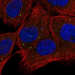
- Experimental details
- Immunofluorescent staining of human cell line A-431 shows positivity in vesicles.
- Sample type
- Human

Supportive validation
- Submitted by
- Atlas Antibodies (provider)
- Enhanced method
- Orthogonal validation
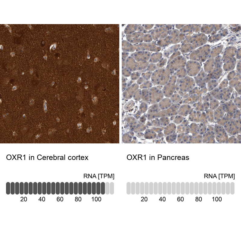
- Main image

- Experimental details
- Immunohistochemistry analysis in human cerebral cortex and pancreas tissues using HPA027395 antibody. Corresponding OXR1 RNA-seq data are presented for the same tissues.
- Sample type
- Human
- Protocol
- Protocol