Explore
 Validate
 Learn
Learn Western blot
 Immunohistochemistry
Antibody data
- Antibody Data
- Antigen structure
- References [1]
- Comments [0]
- Validations
- Immunohistochemistry [1]
- Other assay [1]
Submit
Validation data
Reference
Comment
Report error
- Product number
- PA5-31746 - Provider product page

- Provider
- Invitrogen Antibodies
- Product name
- OXR1 Polyclonal Antibody
- Antibody type
- Polyclonal
- Antigen
- Recombinant full-length protein
- Description
- Recommended positive controls: NT2D1, IMR32, U87-MG, MCF-7. Predicted reactivity: Mouse (90%), Rat (89%). Store product as a concentrated solution. Centrifuge briefly prior to opening the vial.
- Reactivity
- Human
- Host
- Rabbit
- Isotype
- IgG
- Vial size
- 100 μL
- Concentration
- 1 mg/mL
- Storage
- Store at 4°C short term. For long term storage, store at -20°C, avoiding freeze/thaw cycles.
Submitted references Mapping the H(+) (V)-ATPase interactome: identification of proteins involved in trafficking, folding, assembly and phosphorylation.
Merkulova M, Păunescu TG, Azroyan A, Marshansky V, Breton S, Brown D
Scientific reports 2015 Oct 7;5:14827
Scientific reports 2015 Oct 7;5:14827
No comments: Submit comment
Supportive validation
- Submitted by
- Invitrogen Antibodies (provider)
- Main image
- Experimental details
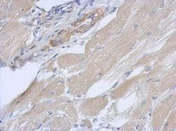
- Immunohistochemical analysis of paraffin-embedded human gastric cancer, using OXR1 (Product # PA5-31746) antibody at 1:500 dilution. Antigen Retrieval: EDTA based buffer, pH 8.0, 15 min.

Supportive validation
- Submitted by
- Invitrogen Antibodies (provider)
- Main image
- Experimental details
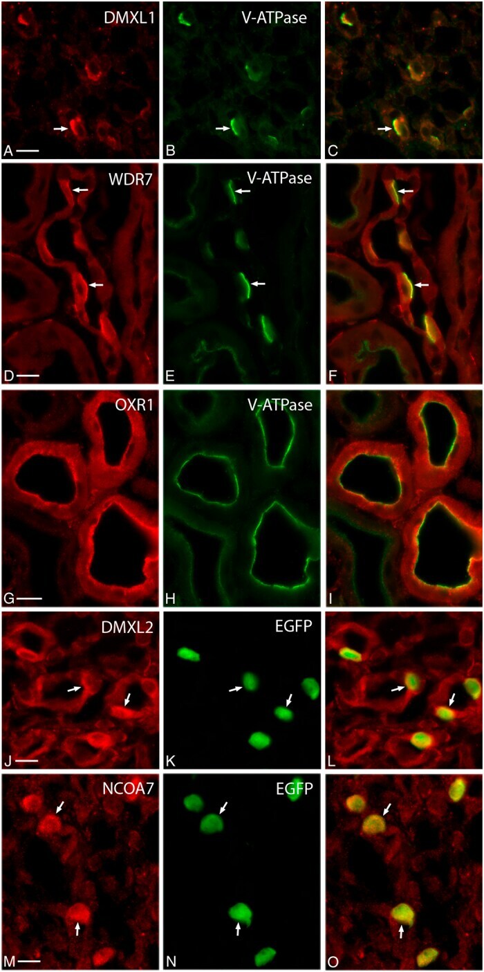
- Figure 2 Localization of DMXL1, DMXL2, NCOA7, OXR1 and WDR7 in proton secreting cells of mouse kidney by immunocytochemistry. DMXL1 (( A) , red) co-localizes with the A-subunit of V-ATPase (( B ), green) in intercalated cells (ICs, indicated with arrows) of wild type mouse inner medullary collecting duct. WDR7 (( D ), red) also co-localizes with the same V-ATPase subunit ( E ), green) in ICs of wild type mouse cortical collecting duct. OXR1 ( G ), red) and V-ATPase (( H ), green) co-localize in the apical pole of distal convoluted tubule cells of wild type mice. DMXL2 ( J ), red) and NCOA7 ( M ), red) are expressed predominantly in ICs of B1-EGFP transgenic mouse inner medullary collecting duct (ICs are green in panels ( K , N )). Note, that in the B1-EGFP transgenic mouse EGFP is not fused to B1 subunit, rather EGFP expression is driven by B1 promoter. Thus, while EGFP is expressed specifically in ICs, its localization in these cells is purely cytosolic and does not follow V-ATPase sub-cellular localization pattern. Merged images ( C,F,I,L,O ) are shown in the right column. Scale bar = 10 mum.