Explore
 Validate
 Learn
Learn Western blot
Antibody data
- Antibody Data
- Antigen structure
- References [0]
- Comments [0]
- Validations
- Western blot [1]
- Immunohistochemistry [2]
Submit
Validation data
Reference
Comment
Report error
- Product number
- ASR-081-200UL - Provider product page

- Provider
- Invitrogen Antibodies
- Product name
- Slitrk1 (extracellular) Polyclonal Antibody
- Antibody type
- Polyclonal
- Antigen
- Other
- Reactivity
- Human, Mouse, Rat
- Host
- Rabbit
- Isotype
- IgG
- Vial size
- 200 µL
- Concentration
- 0.8 mg/mL
- Storage
- -20° C, Avoid Freeze/Thaw Cycles
No comments: Submit comment
Supportive validation
- Submitted by
- Invitrogen Antibodies (provider)
- Main image
- Experimental details
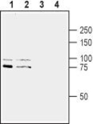
- Western blot analysis of rat brain lysate (lanes 1 and 3) and mouse brain synaptosomal fraction (lanes 2 and 4): - 1,2. Anti-Slitrk1 (extracellular) Antibody (#ASR-081), (1:200).3,4. Anti-Slitrk1 (extracellular) Antibody , preincubated with Slitrk1 (extracellular) Blocking Peptide (#BLP-SR081).

Supportive validation
- Submitted by
- Invitrogen Antibodies (provider)
- Main image
- Experimental details
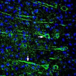
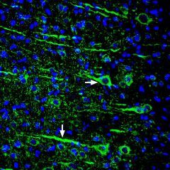
- Expression of Slitrk1 in rat cortex - Immunohistochemical staining of perfusion-fixed frozen rat brain sections using Anti-Slitrk1 (extracellular) Antibody (#ASR-081), (1:300), followed by goat Anti-rabbit-AlexaFluor-488.Slitrk1staining (green) in the rat parieto-temporal cortex is detected in pyramidal neurons (horizontal arrow) and their dendrites (vertical arrow). Cell nuclei are stained with DAPI (blue).
- Submitted by
- Invitrogen Antibodies (provider)
- Main image
- Experimental details
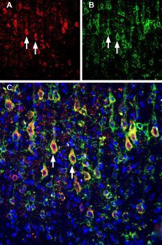
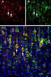
- Multiplex staining of Slitrk1 and KV2.1 in rat parietal cortex - Immunohistochemical staining of immersion-fixed, free floating rat brain frozen sections using rabbit Anti-Slitrk1 (extracellular) Antibody (#ASR-081), (1:200) andGuinea pig Anti-KV2.1 Antibody (#APC-012-GP), (1:200). A. Slitrk1 staining (red) appears in profiles of pyramidal neurons. B. KV2.1 staining (green) is detected in profiles of pyramidal neurons. C. Merge of the two images shows colocalization in several neurons (arrows). Cell nuclei are stained with DAPI (blue).