 Explore
 Validate
 Learn
Learn Western blot
 ELISA
ELISAAntibody data
- Antibody Data
- Antigen structure
- References [0]
- Comments [0]
- Validations
- Western blot [3]
- Immunocytochemistry [2]
- Immunohistochemistry [1]
Submit
Validation data
Reference
Comment
Report error
- Product number
- PA5-98497 - Provider product page

- Provider
- Invitrogen Antibodies
- Product name
- SPOCK1 Polyclonal Antibody
- Antibody type
- Polyclonal
- Antigen
- Recombinant full-length protein
- Reactivity
- Human, Mouse
- Host
- Rabbit
- Isotype
- IgG
- Vial size
- 100 μg
- Concentration
- 1 mg/mL
- Storage
- -20°C or -80°C if preferred
No comments: Submit comment
Supportive validation
- Submitted by
- Invitrogen Antibodies (provider)
- Main image

- Experimental details
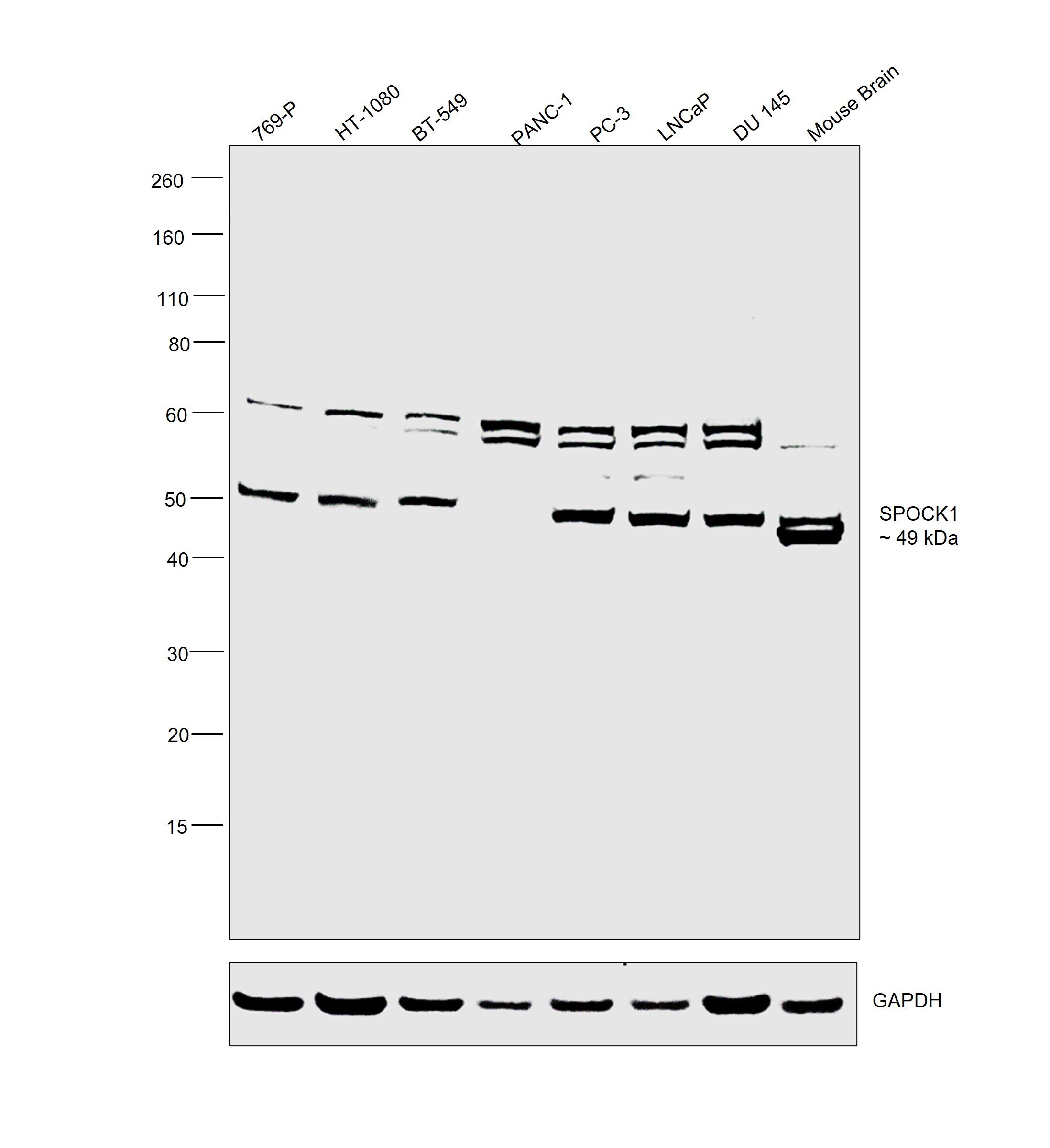
- Western blot was performed using SPOCK1 Polyclonal Antibody (Product # PA5-98497) and a 49 kDa band corresponding to SPOCK1 was observed across cell lines and tissue tested. Whole cell extracts (30 µg lysate) of 769-P (Lane 1), HT-1080 (Lane 2), BT-549 (Lane 3), PANC-1 (Lane 4), PC-3 (Lane 5), LNCaP (Lane 6), DU 145 (Lane 7), Mouse Brain (Lane 8) were electrophoresed using NuPAGE™ 4-12% Bis-Tris Protein Gel (Product # NP0322BOX), 12 well. Resolved proteins were then transferred onto a nitrocellulose membrane (Product # IB23001) by iBlot® 2 Dry Blotting System (Product # IB21001). The blot was probed with the primary antibody (1:2000 dilution) and detected by chemiluminescence with Goat anti-Rabbit IgG (Heavy Chain) Superclonal™ Recombinant Secondary Antibody, HRP (Product # A27036, 1:20,000 dilution) using the iBright™ FL1500 Imaging System (Product # A44115). Chemiluminescent detection was performed using SuperSignal™ West Atto Ultimate Sensitivity Substrate (Product # A38556).There was a non specific band observed at 63 kDa. SPOCK-1 was observed to be at low levels in PANC-1 as reported.
- Submitted by
- Invitrogen Antibodies (provider)
- Main image
- Experimental details
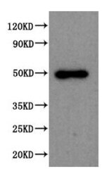
- Western Blot analysis of SPOCK1 using a SPOCK1 Polyclonal antibody (Product # PA5-98497). Positive WB detected in: 293T whole cell lysate. All lanes: SPOCK1 antibody at 1:2000. A secondary Goat polyclonal antibody to rabbit IgG was applied at a 1:50,000 dilution. Observed band size: 50 kDa.

- Submitted by
- Invitrogen Antibodies (provider)
- Main image
- Experimental details
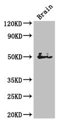
- Western Blot analysis of SPOCK1 using a SPOCK1 Polyclonal antibody (Product # PA5-98497). Positive WB detected in: Mouse brain tissue. All lanes: SPOCK1 antibody at 1:3000. A secondary Goat polyclonal antibody to rabbit IgG was applied at a 1:50,000 dilution. Observed band size: 50 kDa.

Supportive validation
- Submitted by
- Invitrogen Antibodies (provider)
- Main image

- Experimental details
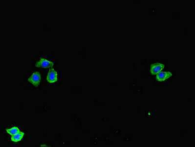
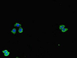
- Immunofluorescent analysis of SPOCK1 in HepG2 cells using a SPOCK1 polyclonal antibody (Product # PA5-98497) at a dilution of 1:100. Alexa Fluor 488-congugated Goat Anti-Rabbit IgG(H+L) secondary antibody was used.
- Submitted by
- Invitrogen Antibodies (provider)
- Main image
- Experimental details
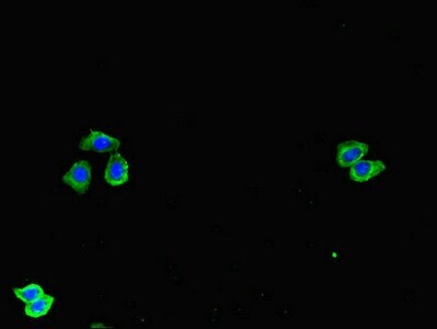
- Immunofluorescent analysis of SPOCK1 in HepG2 cells using a SPOCK1 polyclonal antibody (Product # PA5-98497) at a dilution of 1:100. Alexa Fluor 488-congugated Goat Anti-Rabbit IgG(H+L) secondary antibody was used.
Supportive validation
- Submitted by
- Invitrogen Antibodies (provider)
- Main image
- Experimental details
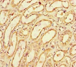
- Immunohistochemical analysis of SPOCK1 in paraffin embedded human kidney tissue using a SPOCK1 polyclonal antibody (Product # PA5-98497) at a dilution of 1:100.
