Explore
 Validate
 Learn
Learn Western blot
Antibody data
- Antibody Data
- Antigen structure
- References [0]
- Comments [0]
- Validations
- Western blot [1]
- Immunocytochemistry [1]
- Flow cytometry [1]
Submit
Validation data
Reference
Comment
Report error
- Product number
- MAB8559 - Provider product page

- Provider
- R&D Systems
- Product name
- Human IGDCC3 Antibody
- Antibody type
- Monoclonal
- Description
- Protein A or G purified from hybridoma culture supernatant. Detects human IGDCC3 in direct ELISAs.
- Reactivity
- Human
- Host
- Mouse
- Conjugate
- Unconjugated
- Antigen sequence
Q81VU1- Isotype
- IgG
- Antibody clone number
- 920038
- Vial size
- 100 ug
- Storage
- Use a manual defrost freezer and avoid repeated freeze-thaw cycles. 12 months from date of receipt, -20 to -70 °C as supplied. 1 month, 2 to 8 °C under sterile conditions after reconstitution. 6 months, -20 to -70 °C under sterile conditions after reconstitution.
No comments: Submit comment
Supportive validation
- Submitted by
- R&D Systems (provider)
- Main image
- Experimental details
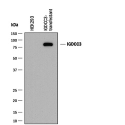
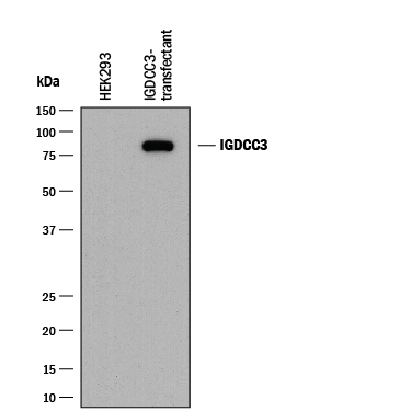
- Detection of Human IGDCC3 by Western Blot. Western blot shows lysates of HEK293 human embryonic kidney cell line either mock-transfected or transfected with human IGDCC3. PVDF membrane was probed with 2 µg/mL of Mouse Anti-Human IGDCC3 Monoclonal Antibody (Catalog # MAB8559) followed by HRP-conjugated Anti-Mouse IgG Secondary Antibody (Catalog # HAF018). A specific band was detected for IGDCC3 at approximately 85 kDa (as indicated). This experiment was conducted under reducing conditions and using Immunoblot Buffer Group 1.
Supportive validation
- Submitted by
- R&D Systems (provider)
- Main image
- Experimental details
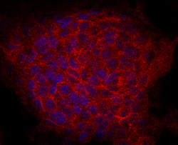
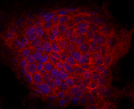
- IGDCC3 in HepG2 Human Cell Line. IGDCC3 was detected in immersion fixed HepG2 human hepatocellular carcinoma cell line using Mouse Anti-Human IGDCC3 Monoclonal Antibody (Catalog # MAB8559) at 10 µg/mL for 3 hours at room temperature. Cells were stained using the NorthernLights™ 557-conjugated Anti-Mouse IgG Secondary Antibody (red; Catalog # NL007) and counterstained with DAPI (blue). Specific staining was localized to cytoplasm and cell surfaces. View our protocol for Fluorescent ICC Staining of Cells on Coverslips.
Supportive validation
- Submitted by
- R&D Systems (provider)
- Main image
- Experimental details
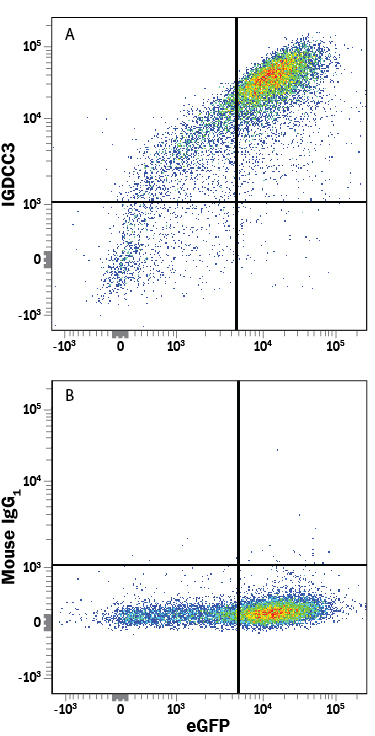
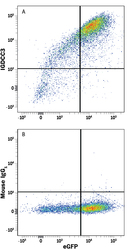
- Detection of IGDCC3 in HEK293 Human Cell Line Transfected with Human IGDCC3 and eGFP by Flow Cytometry. HEK293 human embryonic kidney cell line transfected with human IGDCC3 and eGFP was stained with either (A) Mouse Anti-Human IGDCC3 Monoclonal Antibody (Catalog # MAB8559) or (B) Mouse IgG1 Isotype Control (Catalog # MAB002) followed by Allophycocyanin-conjugated Anti-Mouse IgG Secondary Antibody (Catalog # F0101B).