Explore
 Validate
 Learn
Learn Western blot
 Immunocytochemistry
 Immunohistochemistry
Antibody data
- Antibody Data
- Antigen structure
- References [1]
- Comments [0]
- Validations
- Immunocytochemistry [3]
- Other assay [5]
Submit
Validation data
Reference
Comment
Report error
- Product number
- PA5-103481 - Provider product page

- Provider
- Invitrogen Antibodies
- Product name
- WNT8A Polyclonal Antibody
- Antibody type
- Polyclonal
- Antigen
- Synthetic peptide
- Description
- Antibody detects endogenous levels of total WNT8A.
- Reactivity
- Human, Mouse
- Host
- Rabbit
- Isotype
- IgG
- Vial size
- 100 μL
- Concentration
- 1 mg/mL
- Storage
- -20°C
Submitted references lncRNA Ttc3-209 Promotes the Apoptosis of Retinal Ganglion Cells in Retinal Ischemia Reperfusion Injury by Targeting the miR-484/Wnt8a Axis.
Zhang R, Feng Y, Lu J, Ge Y, Li H
Investigative ophthalmology & visual science 2021 Mar 1;62(3):13
Investigative ophthalmology & visual science 2021 Mar 1;62(3):13
No comments: Submit comment
Supportive validation
- Submitted by
- Invitrogen Antibodies (provider)
- Main image
- Experimental details
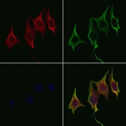
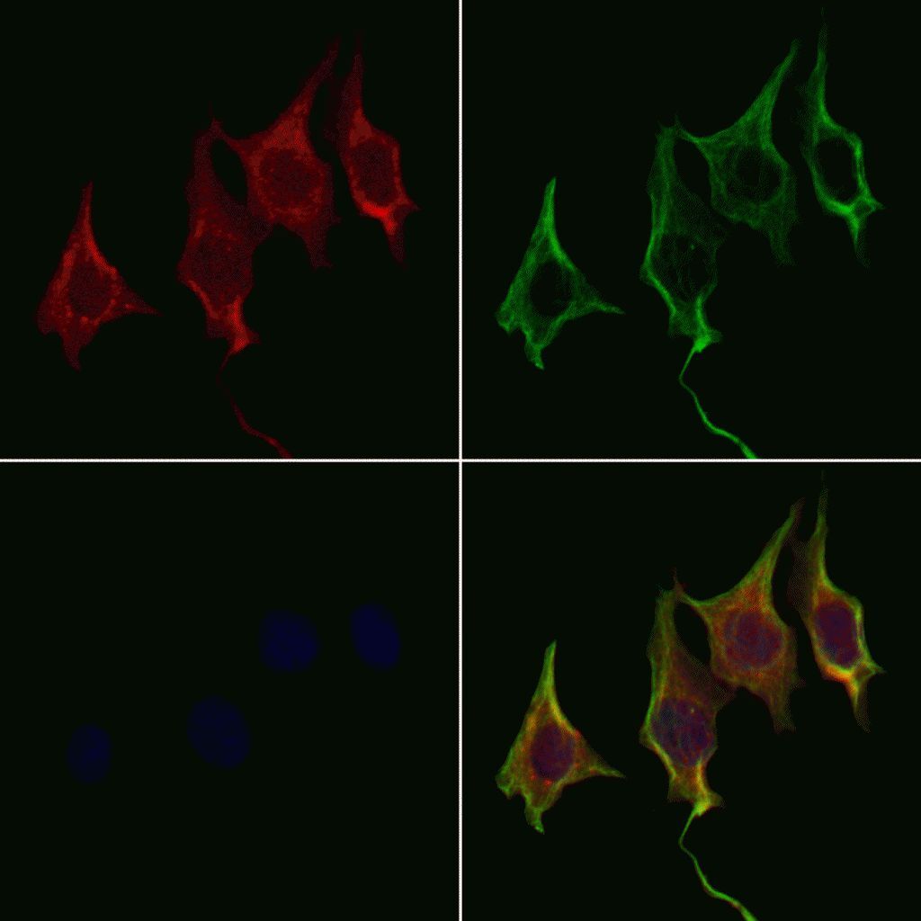
- Immunofluorescent analysis of WNT8A in 3T3 cells. Samples were fixed with paraformaldehyde, permeabilized with 0.1% Triton X-100, blocked with 10% serum (45 min at 25°C), incubated with mouse anti-beta tubulin and WNT8A polyclonal antibody (Product # PA5-103481) using a dilution of 1:200 (1 hr, 37°C), and followed by goat anti-rabbit IgG Alexa Fluor 594 (red) and goat anti-mouse IgG Alexa Fluor 488 (green).

- Submitted by
- Invitrogen Antibodies (provider)
- Main image
- Experimental details
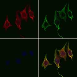
- Immunofluorescent analysis of WNT8A in 3T3 cells. Samples were fixed with paraformaldehyde, permeabilized with 0.1% Triton X-100, blocked with 10% serum (45 min at 25°C), incubated with mouse anti-beta tubulin and WNT8A polyclonal antibody (Product # PA5-103481) using a dilution of 1:200 (1 hr, 37°C), and followed by goat anti-rabbit IgG Alexa Fluor 594 (red) and goat anti-mouse IgG Alexa Fluor 488 (green).
- Submitted by
- Invitrogen Antibodies (provider)
- Main image

- Experimental details
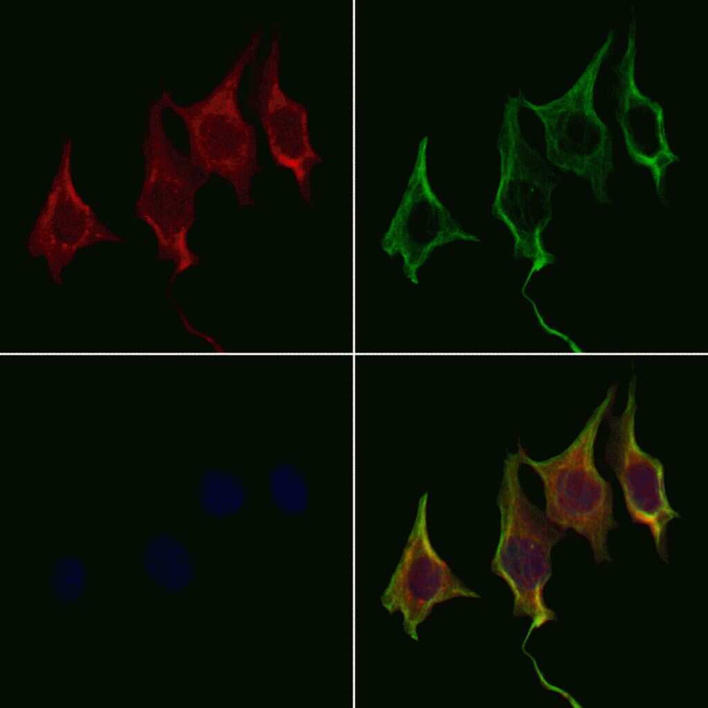
- Immunofluorescent analysis of WNT8A in 3T3 cells. Samples were fixed with paraformaldehyde, permeabilized with 0.1% Triton X-100, blocked with 10% serum (45 min at 25°C), incubated with mouse anti-beta tubulin and WNT8A polyclonal antibody (Product # PA5-103481) using a dilution of 1:200 (1 hr, 37°C), and followed by goat anti-rabbit IgG Alexa Fluor 594 (red) and goat anti-mouse IgG Alexa Fluor 488 (green).
Supportive validation
- Submitted by
- Invitrogen Antibodies (provider)
- Main image
- Experimental details
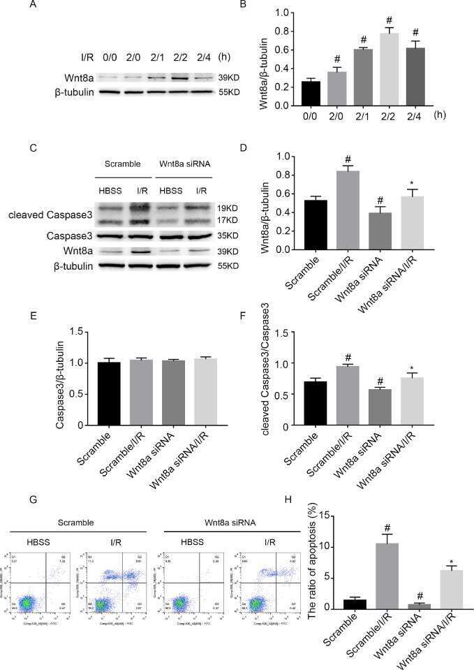
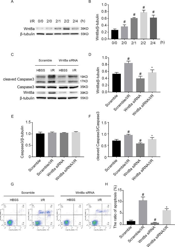
- Figure 7. Wnt8a knockdown suppressed the I/R-induced RGC apoptosis. RGCs were transfected with 50 nmol/L Wnt8a siRNA or scramble and then treated with or without I/R damage. ( A ) RGCs were treated with or without I/R at indicated time points (I/R for 0/0, 2/0, 2/1, 2/2, 2/4 hours, respectively). The expression levels of Wnt8a were detected by Western blot. ( B ) Densitometric measurement of Western blot bands for Wnt8a. ( C ) Western Blot analysis of Wnt8a, caspase-3, and cleaved caspase-3. ( D - F ) Densitometric measurement of Western blot bands for Wnt8a, caspase-3, and cleaved caspase-3. ( G , H ) Flow cytometry analysis of total cell apoptosis. The cells in the Q1 region represent necrotic cells, the cells in the Q2 region represent late apoptotic cells, the cells in the Q3 region represent early apoptotic cells, and the cells in the Q4 region represent normal cells. Data are expressed as mean +- SD ( n = 5). # P < 0.05, I/R for 2/0, 2/1, 2/2, 2/4 hours, respectively, versus 0/0 hour, scramble with I/R group or Wnt8a siRNA group versus scramble group; * P < 0.05, Wnt8a siRNA with I/R group versus scramble with I/R group.
- Submitted by
- Invitrogen Antibodies (provider)
- Main image
- Experimental details
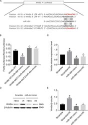
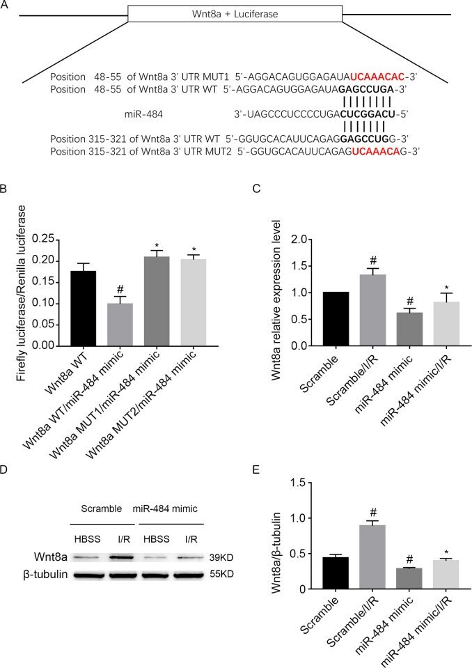
- Figure 6. Wnt8a was identified as a target gene for miR-484. RGCs were transfected with 50 nmol/L miR-484 mimic or scramble and then treated with or without I/R damage. ( A ) Putative miR-484 complementary binding sites in the 3''-UTR of mouse Wnt8a mRNA. ( B ) Detection of luciferase activities after cotransfection with the 3''-UTR luciferase reporter vectors for mouse Wnt8a-WT, Wnt8a-MUT1, Wnt8a-MUT2, miR-484 mimic, or scramble. ( C ) Real-time qPCR analysis of Wnt8a mRNA expression. ( D , E ) Western blotting analysis of Wnt8a ( D ) and densitometric measurement of Western blotting bands ( E ). Data are expressed as mean +- SD ( n = 5). # P < 0.05, Wnt8a-WT/miR-484 mimic group versus Wnt8a-WT group, scramble with I/R group or miR-484 mimic group versus scramble group; * P < 0.05, Wnt8a-MUT1/miR-484 mimic group or Wnt8a-MUT2/miR-484 mimic group versus Wnt8a-WT/miR-484 mimic group, miR-484 mimic with I/R group versus scramble with I/R group.
- Submitted by
- Invitrogen Antibodies (provider)
- Main image

- Experimental details
- Figure 7. Wnt8a knockdown suppressed the I/R-induced RGC apoptosis. RGCs were transfected with 50 nmol/L Wnt8a siRNA or scramble and then treated with or without I/R damage. ( A ) RGCs were treated with or without I/R at indicated time points (I/R for 0/0, 2/0, 2/1, 2/2, 2/4 hours, respectively). The expression levels of Wnt8a were detected by Western blot. ( B ) Densitometric measurement of Western blot bands for Wnt8a. ( C ) Western Blot analysis of Wnt8a, caspase-3, and cleaved caspase-3. ( D - F ) Densitometric measurement of Western blot bands for Wnt8a, caspase-3, and cleaved caspase-3. ( G , H ) Flow cytometry analysis of total cell apoptosis. The cells in the Q1 region represent necrotic cells, the cells in the Q2 region represent late apoptotic cells, the cells in the Q3 region represent early apoptotic cells, and the cells in the Q4 region represent normal cells. Data are expressed as mean +- SD ( n = 5). # P < 0.05, I/R for 2/0, 2/1, 2/2, 2/4 hours, respectively, versus 0/0 hour, scramble with I/R group or Wnt8a siRNA group versus scramble group; * P < 0.05, Wnt8a siRNA with I/R group versus scramble with I/R group.
- Submitted by
- Invitrogen Antibodies (provider)
- Main image
- Experimental details
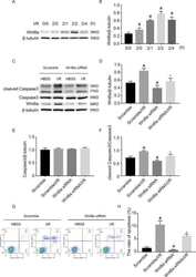
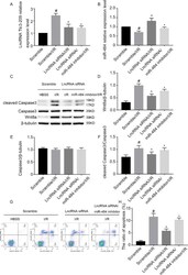
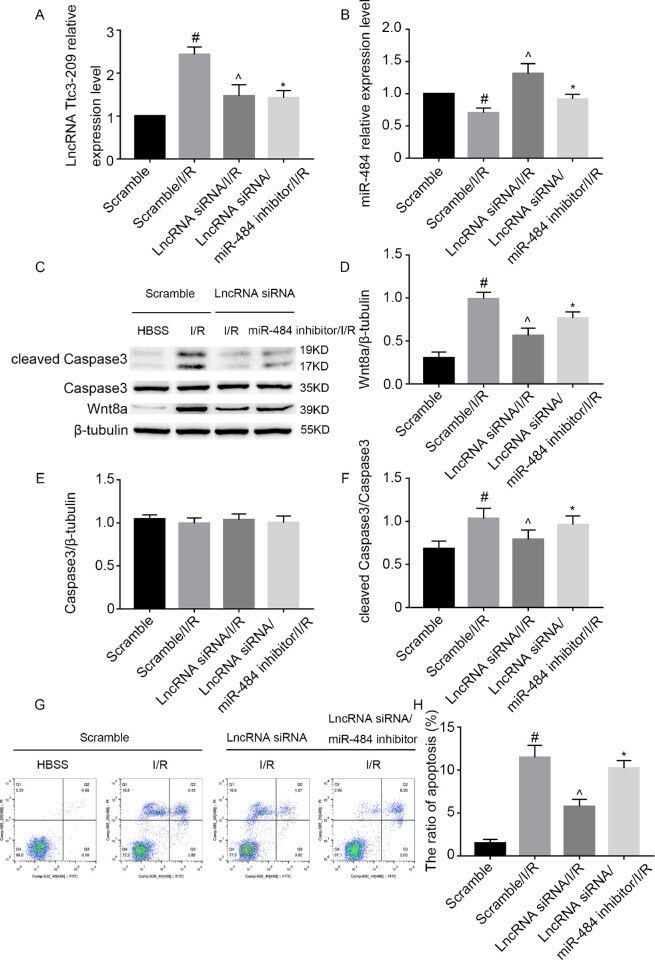
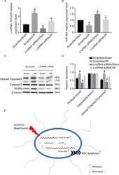
- Figure 8. Inhibition of lncRNA Ttc3-209 alleviated the I/R-induced RGC apoptosis, which was reversed by the inhibitor of miR-484. RGCs were cotransfected with lncRNA Ttc3-209 siRNA (50 nmol/L) and miR-484 inhibitor (50 nmol/L) or scramble and then treated with or without I/R damage. ( A , B ) Real-time qPCR analysis of lncRNA Ttc3-209 expression ( A ) and miR-484 ( B ). ( C - F ) Western blot analysis of Wnt8a, caspase-3, and cleaved caspase-3 ( C ) and densitometric measurement of Western blot bands ( D - F ). ( G , H ) Flow cytometry analysis of total cell apoptosis. The cells in the Q1 region represent necrotic cells, the cells in the Q2 region represent late apoptotic cells, the cells in the Q3 region represent early apoptotic cells, and the cells in the Q4 region represent normal cells. Data are expressed as mean +- SD ( n = 5). # P < 0.05, scramble with I/R group versus scramble group; ^ P < 0.05, lncRNA Ttc3-209 siRNA with I/R group versus scramble with I/R group; * P < 0.05, lncRNA Ttc3-209 siRNA/miR-484 inhibitor with I/R group versus lncRNA Ttc3-209 siRNA with I/R group.
- Submitted by
- Invitrogen Antibodies (provider)
- Main image
- Experimental details
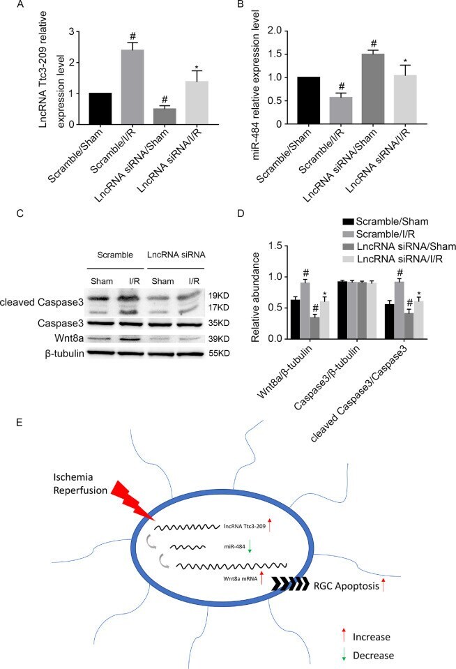
- Figure 10. The downregulation of lncRNA Ttc3-209 diminished I/R-induced apoptosis in RGC apoptosis via the lncRNA Ttc3-209/miR-484/Wnt8a axis in C57BL/6J mice. C57BL/6J mice were preinjected with 1 uL (1 ug/uL) lncRNA Ttc3-209 siRNA or scramble into the vitreous chamber for 24 hours. Next, the intraocular pressure was elevated to 120 mm Hg for 1 hour and then changed to reperfusion for 24 hours. Finally, the retinal tissue was removed to conduct a series of experiments. ( A ) Real-time qPCR analysis of the expression of lncRNA Ttc3-209. ( B ) Real-time qPCR analysis of miR-484 expression. ( C , D ) Western blotting analysis of Wnt8a, caspase-3, and cleaved caspase-3 ( C ) and the densitometric measurement of Western blotting bands ( D ). ( E ) The role and molecular mechanism that regulates the effects of lncRNA Ttc3-209 in the I/R-induced apoptosis of RGCs. Data are expressed as mean +- SD ( n = 5). # P < 0.05, scramble/I/R group or lncRNA Ttc3-209 siRNA/sham group versus scramble/sham group; * P < 0.05, lncRNA Ttc3-209/I/R group versus scramble/I/R group.