Explore
 Validate
 Learn
Learn Western blot
 ELISA
ELISAAntibody data
- Antibody Data
- Antigen structure
- References [7]
- Comments [0]
- Validations
- ELISA [2]
- Immunocytochemistry [2]
- Immunohistochemistry [5]
- Other assay [6]
Submit
Validation data
Reference
Comment
Report error
- Product number
- PA5-27338 - Provider product page

- Provider
- Invitrogen Antibodies
- Product name
- HMOX1 Polyclonal Antibody
- Antibody type
- Polyclonal
- Antigen
- Recombinant full-length protein
- Description
- Recommended positive controls: HeLa, Raw264.7, C2C12, rat spleen, HeLa (500 µM CoCl2 treatment for 24 hr). Predicted reactivity: Mouse (81%), Rat (80%), Dog (85%), Pig (82%), Bovine (83%). Store product as a concentrated solution. Centrifuge briefly prior to opening the vial.
- Reactivity
- Human, Mouse, Rat
- Host
- Rabbit
- Isotype
- IgG
- Vial size
- 100 μL
- Concentration
- 0.54 mg/mL
- Storage
- Store at 4°C short term. For long term storage, store at -20°C, avoiding freeze/thaw cycles.
Submitted references Preventive Effect of Limosilactobacillus fermentum SCHY34 on Lead Acetate-Induced Neurological Damage in SD Rats.
Chronic Hematuria Increases Chronic Kidney Injury and Epithelial-Mesenchymal Transition in 5/6 Nephrectomy Rats.
α-Lipoic Acid Maintains Brain Glucose Metabolism via BDNF/TrkB/HIF-1α Signaling Pathway in P301S Mice.
Defense and protection mechanisms in lung exposed to asbestiform fiber: the role of macrophage migration inhibitory factor and heme oxygenase-1.
Physalis peruviana L. inhibits ovalbumin‑induced airway inflammation by attenuating the activation of NF‑κB and inflammatory molecules.
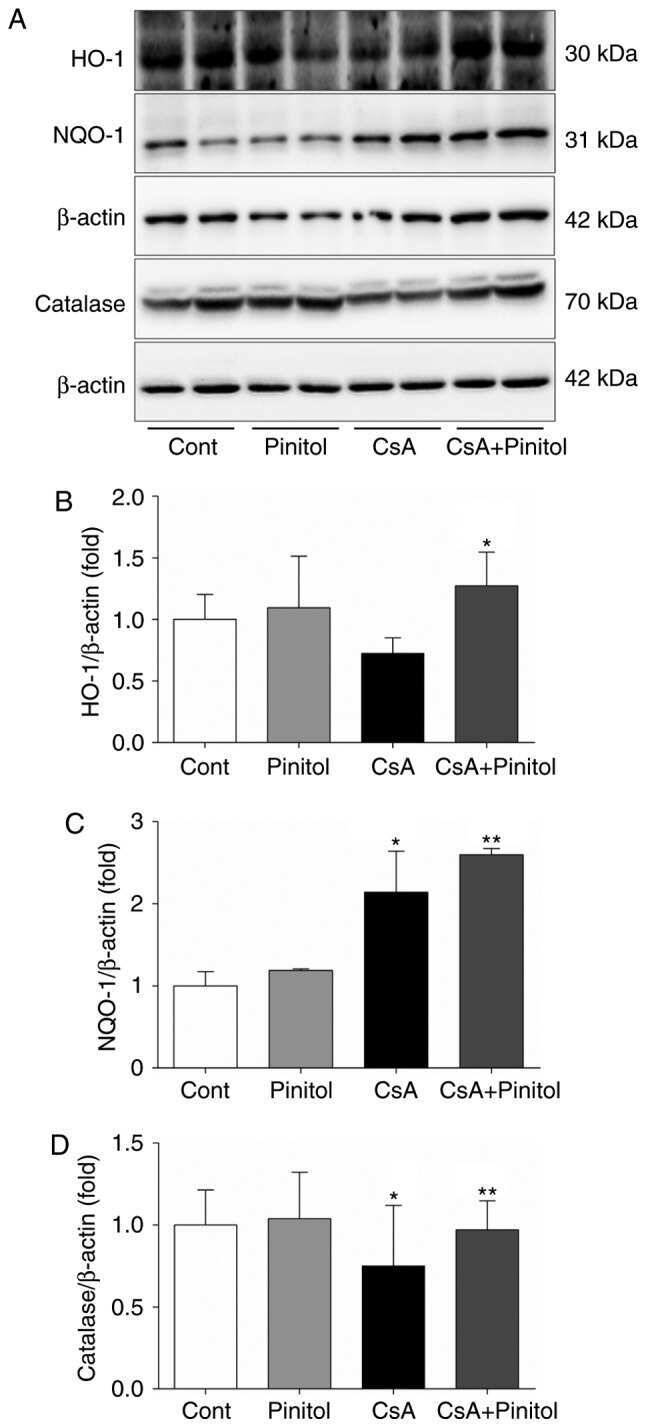
D‑Pinitol alleviates cyclosporine A‑induced renal tubulointerstitial fibrosis via activating Sirt1 and Nrf2 antioxidant pathways.
Hemophagocytosis-mediated keratinization in oral carcinoma in situ and squamous cell carcinoma: a possible histopathogenesis of keratin pearls.
Long X, Wu H, Zhou Y, Wan Y, Kan X, Gong J, Zhao X
Frontiers in nutrition 2022;9:852012
Frontiers in nutrition 2022;9:852012
Chronic Hematuria Increases Chronic Kidney Injury and Epithelial-Mesenchymal Transition in 5/6 Nephrectomy Rats.
Xiao M, Medipally AK, Biederman L, Satoskar AA, Ivanov I, Rovin BH, Brodsky SV
Frontiers in medicine 2021;8:753506
Frontiers in medicine 2021;8:753506
α-Lipoic Acid Maintains Brain Glucose Metabolism via BDNF/TrkB/HIF-1α Signaling Pathway in P301S Mice.
Zhang YH, Yan XZ, Xu SF, Pang ZQ, Li LB, Yang Y, Fan YG, Wang Z, Yu X, Guo C, Ao Q
Frontiers in aging neuroscience 2020;12:262
Frontiers in aging neuroscience 2020;12:262
Defense and protection mechanisms in lung exposed to asbestiform fiber: the role of macrophage migration inhibitory factor and heme oxygenase-1.
Loreto C, Caltabiano R, Graziano ACE, Castorina S, Lombardo C, Filetti V, Vitale E, Rapisarda G, Cardile V, Ledda C, Rapisarda V
European journal of histochemistry : EJH 2020 Apr 16;64(2)
European journal of histochemistry : EJH 2020 Apr 16;64(2)
Physalis peruviana L. inhibits ovalbumin‑induced airway inflammation by attenuating the activation of NF‑κB and inflammatory molecules.
Park HA, Kwon OK, Ryu HW, Min JH, Park MW, Park MH, Paik JH, Choi S, Paryanto I, Yuniato P, Oh SR, Ahn KS, Lee JW
International journal of molecular medicine 2019 Apr;43(4):1830-1838
International journal of molecular medicine 2019 Apr;43(4):1830-1838
D‑Pinitol alleviates cyclosporine A‑induced renal tubulointerstitial fibrosis via activating Sirt1 and Nrf2 antioxidant pathways.
Koh ES, Kim S, Kim M, Hong YA, Shin SJ, Park CW, Chang YS, Chung S, Kim HS
International journal of molecular medicine 2018 Apr;41(4):1826-1834
International journal of molecular medicine 2018 Apr;41(4):1826-1834
Hemophagocytosis-mediated keratinization in oral carcinoma in situ and squamous cell carcinoma: a possible histopathogenesis of keratin pearls.
Al-Eryani K, Cheng J, Abé T, Yamazaki M, Maruyama S, Tsuneki M, Essa A, Babkair H, Saku T
Journal of cellular physiology 2013 Oct;228(10):1977-88
Journal of cellular physiology 2013 Oct;228(10):1977-88
No comments: Submit comment
Supportive validation
- Submitted by
- Invitrogen Antibodies (provider)
- Main image
- Experimental details
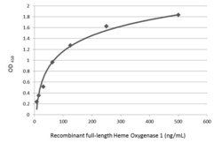
- Sandwich ELISA detection of recombinant full-length Heme Oxygenase 1 protein using HMOX1 Monoclonal Antibody (GT17811) (Product # MA5-31558) as capture antibody at concentration of 5 µg/mL and HMOX1 Polyclonal Antibody (Product # PA5-27338) as detection antibody at concentration of 1 µg/mL. Rabbit IgG antibody (HRP) was diluted at 1:10,000 and used to detect the primary antibody.

- Submitted by
- Invitrogen Antibodies (provider)
- Main image

- Experimental details
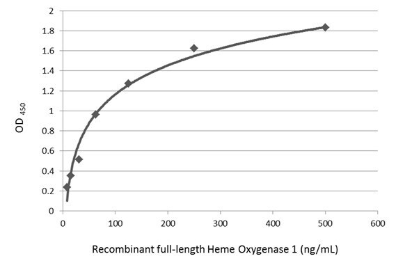
- Sandwich ELISA detection of recombinant full-length Heme Oxygenase 1 protein using HMOX1 Monoclonal Antibody (GT17811) (Product # MA5-31558) as capture antibody at concentration of 5 µg/mL and HMOX1 Polyclonal Antibody (Product # PA5-27338) as detection antibody at concentration of 1 µg/mL. Rabbit IgG antibody (HRP) was diluted at 1:10,000 and used to detect the primary antibody.
Supportive validation
- Submitted by
- Invitrogen Antibodies (provider)
- Main image

- Experimental details
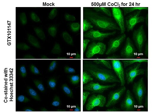
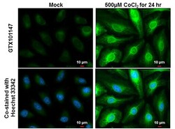
- Immunocytochemistry-Immunofluorescence analysis of HMOX1 was performed in Mock and treated HeLa cells fixed in 4% paraformaldehyde at RT for 15 min. Green: HMOX1 Polyclonal Antibody (Product # PA5-27338) diluted at 1:2000. Blue: Hoechst 33342 staining. Scale bar = 10 µm.
- Submitted by
- Invitrogen Antibodies (provider)
- Main image
- Experimental details
- Immunocytochemistry-Immunofluorescence analysis of HMOX1 was performed in Mock and treated HeLa cells fixed in 4% paraformaldehyde at RT for 15 min. Green: HMOX1 Polyclonal Antibody (Product # PA5-27338) diluted at 1:2000. Blue: Hoechst 33342 staining. Scale bar = 10 µm.

Supportive validation
- Submitted by
- Invitrogen Antibodies (provider)
- Main image
- Experimental details
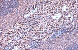
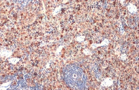
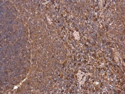
- HMOX1 Polyclonal Antibody detects Heme Oxygenase 1 protein at cytoplasm by immunohistochemical analysis. Sample: Paraffin-embedded rat spleen. Heme Oxygenase 1 stained by HMOX1 Polyclonal Antibody (Product # PA5-27338) diluted at 1:500. Antigen Retrieval: Citrate buffer, pH 6.0, 15 min.
- Submitted by
- Invitrogen Antibodies (provider)
- Main image
- Experimental details
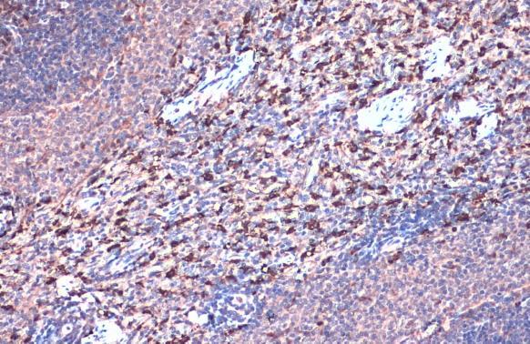
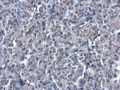
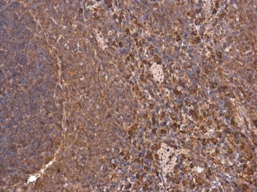
- HMOX1 Polyclonal Antibody detects Heme Oxygenase 1 protein at cytoplasm by immunohistochemical analysis. Sample: Paraffin-embedded mouse spleen. Heme Oxygenase 1 stained by HMOX1 Polyclonal Antibody (Product # PA5-27338) diluted at 1:500. Antigen Retrieval: Citrate buffer, pH 6.0, 15 min.
- Submitted by
- Invitrogen Antibodies (provider)
- Main image
- Experimental details
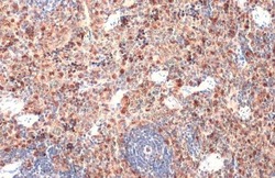
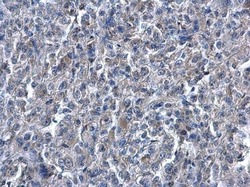
- HMOX1 Polyclonal Antibody detects Heme Oxygenase 1 protein at cytoplasm on human renal carcinoma by immunohistochemical analysis. Sample: Paraffin-embedded human renal carcinoma. HMOX1 Polyclonal Antibody (Product # PA5-27338) diluted at 1:500. Antigen Retrieval: EDTA based buffer, pH 8.0, 15 min.
- Submitted by
- Invitrogen Antibodies (provider)
- Main image
- Experimental details
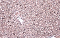
- HMOX1 Polyclonal Antibody detects Heme Oxygenase 1 protein at cytoplasm by immunohistochemical analysis. Sample: Paraffin-embedded mouse liver. Heme Oxygenase 1 stained by HMOX1 Polyclonal Antibody (Product # PA5-27338) diluted at 1:500. Antigen Retrieval: Citrate buffer, pH 6.0, 15 min.

- Submitted by
- Invitrogen Antibodies (provider)
- Main image
- Experimental details
- Immunohistochemistry (Paraffin) analysis of HMOX1 was performed in paraffin-embedded rat spleen tissue using HMOX1 Polyclonal Antibody (Product # PA5-27338) at a dilution of 1:500.
Supportive validation
- Submitted by
- Invitrogen Antibodies (provider)
- Main image
- Experimental details
- NULL
- Submitted by
- Invitrogen Antibodies (provider)
- Main image
- Experimental details
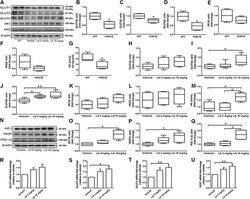
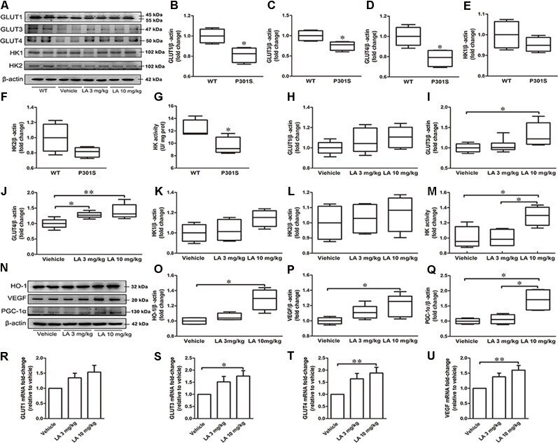
- Figure 1 alpha-Lipoic acid (LA) improved glucose metabolism deficiency in P301S mice. (A) Representative western blots showed the expression levels of glucose transporter 1 (GLUT1), GLUT3, GLUT4, hexokinase-1 (HK-1), and HK2. (B-F) Quantified results of GLUT1, GLUT3, GLUT4, HK1, and HK2 levels between wild type (WT) and P301S mice. beta-actin served as an internal loading control. (G) HK activity between WT and P301S mice. (H-L) Quantified results of the levels of GLUT1, GLUT3, GLUT4, HK-1, and HK2 among vehicle, LA 3 mg/kg, and 10 mg/kg groups. beta-actin served as an internal loading control. (M) HK activity among vehicles, LA 3 mg/kg, and 10 mg/kg groups. (N-Q) Representative western blots and quantified results of the levels of heme oxygenase-1 (HO-1), vascular endothelial growth factor (VEGF), and proliferator-activated receptor gamma coactivator 1-alpha (PGC-1alpha). beta-actin served as an internal loading control. (R-U) mRNA level of GLUT1, GLUT3, GLUT4, and VEGF. Values are represented as the means +- SEM ( n = 7). * p < 0.05, ** p < 0.01.
- Submitted by
- Invitrogen Antibodies (provider)
- Main image
- Experimental details
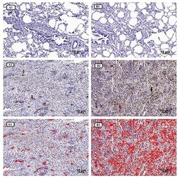
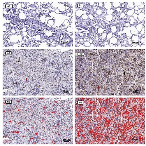
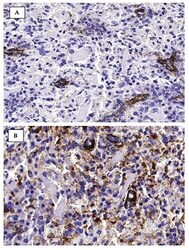
- Figure 1. A1) MIF IHC section of non-exposed sheep (magnification 200x). A2) MIF IHC section of exposed sheep; the fibrotic interstitium showed a strong scattered immunoreaction; green arrow indicates the intra-parenchymal stroma around a bronchiolar structure, yellow arrow shows MIF macrophages immunodetection while red arrow indicates FE deposit fibers (magnification 200x). A3) MIF immunostaining software image analysis of panel A2, in which mainly a high immunostained area (red color) was detected (magnification 200x). B1) HO-1 IHC section of non-exposed sheep (magnification 200x). B2) HO-1 IHC section of exposed sheep; in the lung fibrotic tissue, a strong and widespread immunostaining was demonstrated throughout the interstitium (black arrow) and bronchiolar structures (red arrow) (magnification 200x). B3) HO-1 immunostaining software image analysis of panel B2, in which mainly a high immunostained area (red color) was detected (magnification 200x).
- Submitted by
- Invitrogen Antibodies (provider)
- Main image
- Experimental details
- Figure 2. A) Higher magnification of MIF immunohistochemical expression in exposed sheep section (magnification 400x). B) Higher magnification of HO-1 immunohistochemical expression in exposed sheep section (magnification 400x).

- Submitted by
- Invitrogen Antibodies (provider)
- Main image
- Experimental details
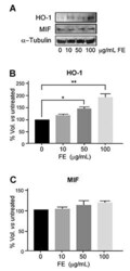
- Figure 4. HO-1 and MIF protein levels in human primary lung fibroblasts unexposed or FE-exposed for 72 h. Representative immunoblotting of HO-1 and MIF expressions (A). Results of three independent immunoblots are represented as percentage of HO-1 (B) and MIF (C) proteins with respect to untreated cells (*P

- Submitted by
- Invitrogen Antibodies (provider)
- Main image

- Experimental details
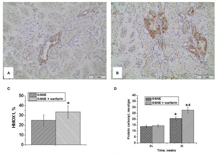
- Figure 4 Expression of heme oxygenase 1 in the kidney and protein carbonyl contents in the renal cortex in kidneys in 5/6 nephrectomy rats with and without warfarin treatment before and at 26 weeks after the surgery. (A) A representative image of the renal cortex from a control 5/6 nephrectomy rat stained an antibody to heme oxygenase 1 (HMOX1). Immunohistochemistry, Magnification 200x. (B) A representative image of the renal cortex from a warfarin-treated 5/6 nephrectomy rat stained an antibody to heme oxygenase 1 (HMOX1). Immunohistochemistry, magnification 200x. (C) Quantitative analysis of HMOX1-positive staining in the tubules, percentage ( n = 6 in 5/6NE control; n = 7 in 5/6NE + warfarin groups). (D) Protein carbonyls were measured in the renal cortex in the right nephrectomy kidney at the time of 5/6 nephrectomy surgery and in the remnant kidney 26 weeks after the surgery. *, p < 0.005 as compared to protein carbonyl contents prior to the surgery; #, p < 0.005 as compared to control 5/6 nephrectomy rats.