Explore
 Validate
 Learn
Learn Western blot
Antibody data
- Antibody Data
- Antigen structure
- References [1]
- Comments [0]
- Validations
- Western blot [2]
Submit
Validation data
Reference
Comment
Report error
- Product number
- MAB3776 - Provider product page

- Provider
- R&D Systems
- Product name
- Human/Mouse HO-1/HMOX1/HSP32 Antibody
- Antibody type
- Monoclonal
- Description
- Protein A or G purified from hybridoma culture supernatant. Detects endogenous human and mouse HO-1 in Western blots. In Western blots, this antibody does not cross-react with rhHO-2.
- Reactivity
- Human, Mouse
- Host
- Rat
- Conjugate
- Unconjugated
- Antigen sequence
P09601- Isotype
- IgG
- Antibody clone number
- 412811
- Vial size
- 100 ug
- Concentration
- LYOPH
- Storage
- Use a manual defrost freezer and avoid repeated freeze-thaw cycles. 12 months from date of receipt, -20 to -70 °C as supplied. 1 month, 2 to 8 °C under sterile conditions after reconstitution. 6 months, -20 to -70 °C under sterile conditions after reconstitution.
Submitted references Induction of heme oxygenase-1 in factor VIII-deficient mice reduces the immune response to therapeutic factor VIII.
Dimitrov JD, Dasgupta S, Navarrete AM, Delignat S, Repesse Y, Meslier Y, Planchais C, Teyssandier M, Motterlini R, Bayry J, Kaveri SV, Lacroix-Desmazes S
Blood 2010 Apr 1;115(13):2682-5
Blood 2010 Apr 1;115(13):2682-5
No comments: Submit comment
Supportive validation
- Submitted by
- R&D Systems (provider)
- Main image
- Experimental details
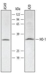
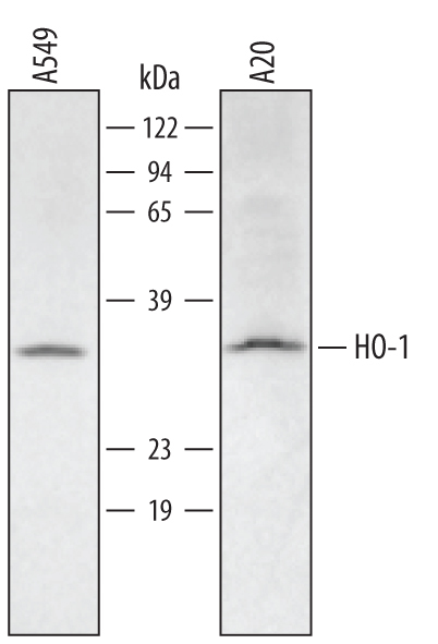
- Detection of Human/Mouse HO-1/HMOX1/HSP32 by Western Blot. Western blot shows lysates of A549 human lung carcinoma cell line and A20 mouse B cell lymphoma cell line. PVDF membrane was probed with 1 µg/mL of Human/Mouse HO-1/HMOX1/HSP32 Monoclonal Antibody (Catalog # MAB3776) followed by HRP-conjugated Anti-Rat IgG Secondary Antibody (Catalog # HAF005). A specific band was detected for HO-1/HMOX1/HSP32 at approximately 32 kDa (as indicated). This experiment was conducted under reducing conditions and using Immunoblot Buffer Group 2.
- Submitted by
- R&D Systems (provider)
- Main image
- Experimental details
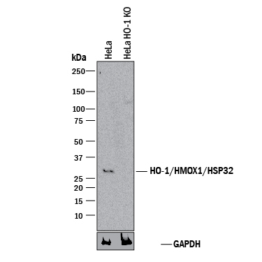
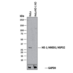
- Western Blot Shows Human HO-1/HMOX1/HSP32 Specificity by Using Knockout Cell Line. Western blot shows lysates of HeLa human cervical epithelial carcinoma parental cell line and HO-1/HMOX1/HSP32 knockout HeLa cell line (KO). PVDF membrane was probed with 1 µg/mL of Rat Anti-Human/Mouse HO-1/HMOX1/HSP32 Monoclonal Antibody (Catalog # MAB3776) followed by HRP-conjugated Anti-Rat IgG Secondary Antibody (Catalog # HAF005). A specific band was detected for HO-1/HMOX1/HSP32 at approximately 32 kDa (as indicated) in the parental HeLa cell line, but is not detectable in knockout HeLa cell line. GAPDH (Catalog # MAB5718) is shown as a loading control. This experiment was conducted under reducing conditions and using Immunoblot Buffer Group 1.