Explore
 Validate
 Learn
Learn Immunocytochemistry
Antibody data
- Antibody Data
- Antigen structure
- References [1]
- Comments [0]
- Validations
- Immunocytochemistry [2]
- Immunohistochemistry [3]
- Other assay [7]
Submit
Validation data
Reference
Comment
Report error
- Product number
- PA5-58204 - Provider product page

- Provider
- Invitrogen Antibodies
- Product name
- METTL14 Polyclonal Antibody
- Antibody type
- Polyclonal
- Antigen
- Recombinant protein fragment
- Description
- Immunogen sequence: RSWNMDSRLQ EIRERQKLRR QLLAQQLGAE SADSIGAVLN SKDEQREIAE TRETCRASYD TSAPNAKRKY LDEGETDEDK MEEYKDELEM QQDEE Highest antigen sequence identity to the following orthologs: Mouse - 92%, Rat - 92%.
- Reactivity
- Human
- Host
- Rabbit
- Isotype
- IgG
- Vial size
- 100 μL
- Concentration
- 0.30 mg/mL
- Storage
- Store at 4°C short term. For long term storage, store at -20°C, avoiding freeze/thaw cycles.
Submitted references Lamin A safeguards the m(6) A methylase METTL14 nuclear speckle reservoir to prevent cellular senescence.
Zhang J, Ao Y, Zhang Z, Mo Y, Peng L, Jiang Y, Wang Z, Liu B
Aging cell 2020 Oct;19(10):e13215
Aging cell 2020 Oct;19(10):e13215
No comments: Submit comment
Supportive validation
- Submitted by
- Invitrogen Antibodies (provider)
- Main image

- Experimental details
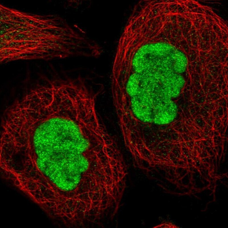
- Immunofluorescent staining of METTL14 in human cell line A-431 shows positivity in nucleus but excluded from the nucleoli. Samples were probed using a METTL14 Polyclonal Antibody (Product # PA5-58204).

- Submitted by
- Invitrogen Antibodies (provider)
- Main image

- Experimental details
- Immunofluorecent analysis of METTL14 in human cell line A-431 using METTL14 Polyclonal Antibody (Product # PA5-58204). Staining shows localization to nucleoplasm.
Supportive validation
- Submitted by
- Invitrogen Antibodies (provider)
- Main image

- Experimental details
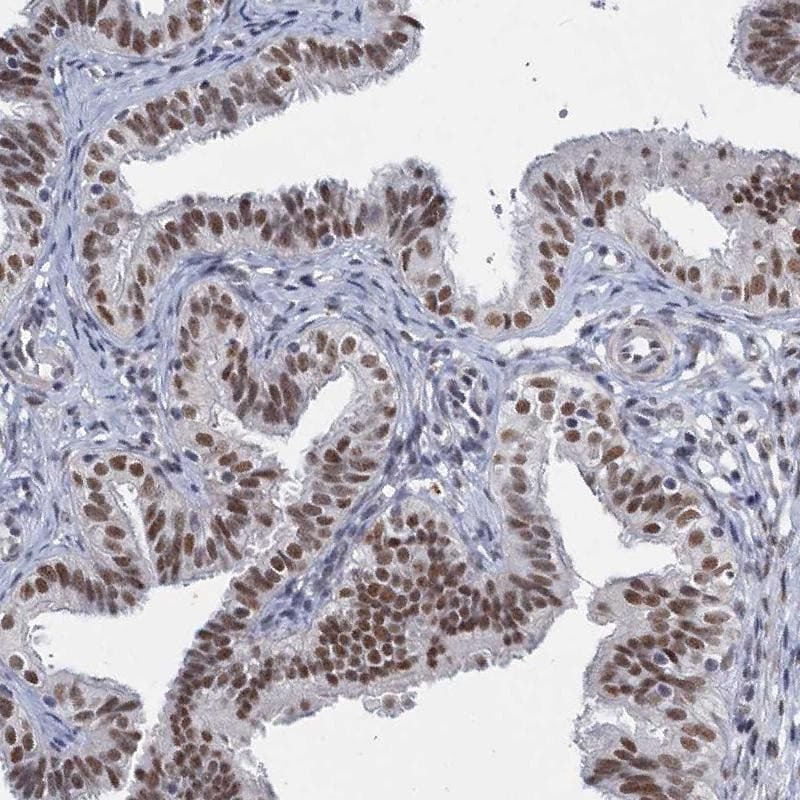
- Immunohistochemical staining of METTL14 in human fallopian tube using a METTL14 Polyclonal Antibody (Product # PA5-58204) shows strong nuclear positivity in glandular cells.
- Submitted by
- Invitrogen Antibodies (provider)
- Main image

- Experimental details
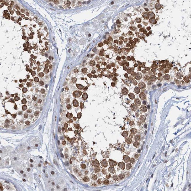
- Immunohistochemical staining of METTL14 in human testis using a METTL14 Polyclonal Antibody (Product # PA5-58204) shows moderate to strong nuclear positivity in cells in seminiferous ducts.
- Submitted by
- Invitrogen Antibodies (provider)
- Main image
- Experimental details
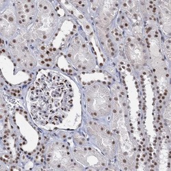
- Immunohistochemical staining of METTL14 in human kidney using a METTL14 Polyclonal Antibody (Product # PA5-58204) shows moderate to strong nuclear positivity in glomeruli and cells in tubules.

Supportive validation
- Submitted by
- Invitrogen Antibodies (provider)
- Main image

- Experimental details
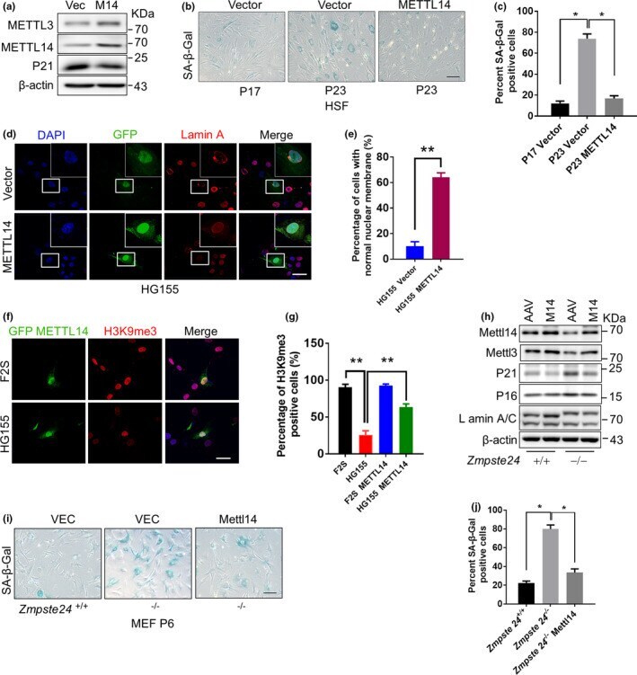
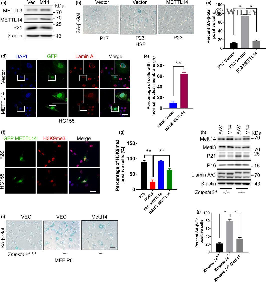
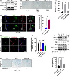
- 5 FIGURE METTL14 overexpression ameliorates senescence. (a) Western blot analysis of METTL3/14 and p21 levels in lenti-METTL14 (lenti-M14)-infected HSFs at P23. (b) Representative images of SA-beta-Gal staining of lenti-M14-infected HSFs at the indicated passages. Scale bar, 100 um. (c) Quantification of the beta-Gal-positive cells in (b) based on 10 randomly chosen views for each group. The data represent the means +- SEM . * p < 0.05. (d) Representative immunofluorescence images of METTL14 and Lamin A expression in lenti-METTL14-infected HG155 at P23. Scale bar, 100 um. (e) The percentage of cells with a normal nuclear shape (%) based on staining from 10 randomly chosen views for each group in (d). The data represent the means +- SEM . ** p < 0.01. (f) Representative immunofluorescence images of METTL14 and H3K9me3 staining in lenti-METTL14-infected HG155 at P23. Scale bar, 100 um. (g) Quantification of (f). The percentage of H3K9me3-positive cells (%) based on 10 randomly chosen views for each group. The data represent the means +- SEM . ** p < 0.01. (h) Western blot analysis of METTL3/14, p21, and p16 levels in adeno-associated virus (AAV)-METTL14-infected Zmpste24 -/- and Zmpste24 +/+ MEFs at P6. (i) SA-beta-Gal staining of AAV-METTL14-infected Zmpste24 -/- MEFs at P6. Scale bar, 100 um. (j) Quantification of the SA-beta-Gal-positive cells in (i) from 10 randomly chosen views for each group. The data represent the means +- SEM . * p < 0.05. The data represent three indep
- Submitted by
- Invitrogen Antibodies (provider)
- Main image
- Experimental details
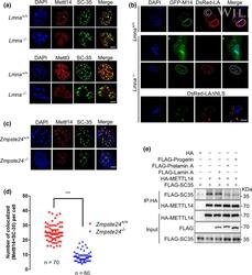
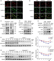
- 2 FIGURE The nuclear localization of METTL3/14 requires Lamin A. (a) Representative immunofluorescence analysis of METTL3, METTL14, and SC35 foci in Lmna +/+ and Lmna -/- MEFs. (b) Representative immunofluorescence analysis of GFP-METTL14 (GFP-M14) in Lmna +/+ and Lmna -/- MEFs with or without DsRed-Lamin A (Red-LA). (c) Representative immunofluorescence analysis of METTL14 and SC35 foci in Zmpste24 +/+ and Zmpste24 -/- MEFs. (d) The quantitative analysis of (c). *** p < 0.001. (e) Co-IP and Western blot analysis of the interaction between SC35 and METTL3/14 in the presence of Lamin A, Progerin, and prelamin A. Scale bars, 10 um

- Submitted by
- Invitrogen Antibodies (provider)
- Main image
- Experimental details
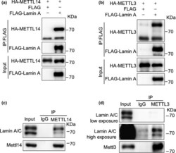
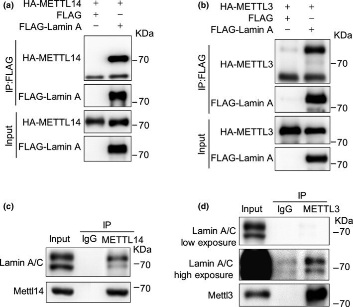
- FIGURE 1 Lamin A interacts with METTL3/14. (a-b) Co-immunoprecipitation (Co-IP) and Western blot analysis of the interactions between FLAG-Lamin A and HA-METTL14 (a); and FLAG-Lamin A HA-METTL3 (b) in HEK293 cells. (c-d) Co-IP and Western blot analysis of the endogenous interaction between Lamin A and METTL14 (c) and Lamin A and METTL3 (d) in MEFs
- Submitted by
- Invitrogen Antibodies (provider)
- Main image

- Experimental details
- FIGURE 2 The nuclear localization of METTL3/14 requires Lamin A. (a) Representative immunofluorescence analysis of METTL3, METTL14, and SC35 foci in Lmna +/+ and Lmna -/- MEFs. (b) Representative immunofluorescence analysis of GFP-METTL14 (GFP-M14) in Lmna +/+ and Lmna -/- MEFs with or without DsRed-Lamin A (Red-LA). (c) Representative immunofluorescence analysis of METTL14 and SC35 foci in Zmpste24 +/+ and Zmpste24 -/- MEFs. (d) The quantitative analysis of (c). *** p < 0.001. (e) Co-IP and Western blot analysis of the interaction between SC35 and METTL3/14 in the presence of Lamin A, Progerin, and prelamin A. Scale bars, 10 um

- Submitted by
- Invitrogen Antibodies (provider)
- Main image
- Experimental details
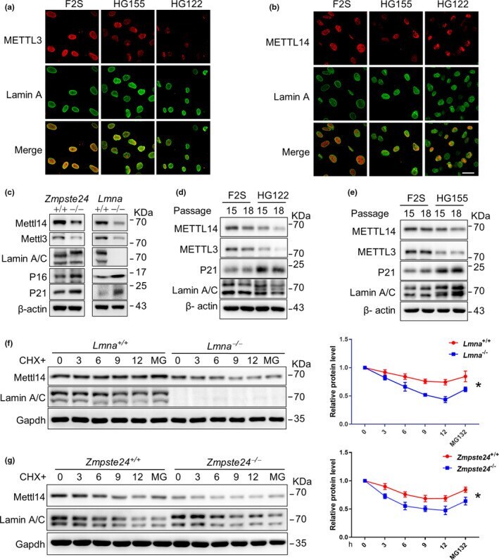
- FIGURE 3 Lamin A abnormality destabilizes METTL14 protein. (a, b) Representative immunofluorescence images showing METTL14/3 and Lamin A expression in skin fibroblasts derived from individual HGPS patients (HG122 and HG155) and normal human skin fibroblasts (F2S). Scale bar, 50 um. (c) Western blot analysis of METTL3/14 protein levels in Zmpste24 +/+ , Zmpste24 -/- , Lmna +/+ , and Lmna -/- MEFs. The data represent three independently derived sets of MEFs in separate experiments. (d, e) Western blot analysis of METTL3/14 protein levels in HG122, HG155, and F2S cells at different passages. (f, g) Western blot analysis of METTL3/14 expression in Lmna -/- (f) and Zmpste24 -/- (g) MEFs compared with WT cells. Cells were treated with cycloheximide (CHX) and/or MG132 (MG) for indicated time. Quantification was performed by Image J (r) . The data represent the means +- SEM . * p < 0.05
- Submitted by
- Invitrogen Antibodies (provider)
- Main image
- Experimental details
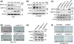
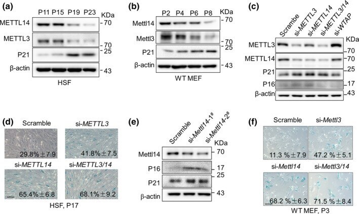
- FIGURE 4 METTL3/14 decline accelerates senescence. (a, b) Western blot analysis of the METTL3/14 and p21 WIF1 levels with subsequent passaging of human skin fibroblasts (HSFs) (a) and wild-type (WT) mouse embryonic fibroblasts (MEFs) (b). (c) Western blot analysis of METTL3/14, p21, and p16 protein levels in HSFs (P17) treated with the indicated siRNAs. (d) Representative images of SA-beta-Gal staining in HSFs (P17) treated with the indicated siRNAs. The percentage of SA-beta-Gal-positive cells is shown. The data represent the means +- SEM . Scale bar, 100 um. (e) Western blot analysis of METTL14, p21, and p16 levels in MEFs (P3) treated with the indicated siRNAs. (f) Representative images of SA-beta-Gal staining in WT MEFs treated with the indicated siRNAs. The percentage of SA-beta-Gal-positive cells is shown. The data represent the means +- SEM . Scale bar, 100 um
- Submitted by
- Invitrogen Antibodies (provider)
- Main image
- Experimental details
- FIGURE 5 METTL14 overexpression ameliorates senescence. (a) Western blot analysis of METTL3/14 and p21 levels in lenti-METTL14 (lenti-M14)-infected HSFs at P23. (b) Representative images of SA-beta-Gal staining of lenti-M14-infected HSFs at the indicated passages. Scale bar, 100 um. (c) Quantification of the beta-Gal-positive cells in (b) based on 10 randomly chosen views for each group. The data represent the means +- SEM . * p < 0.05. (d) Representative immunofluorescence images of METTL14 and Lamin A expression in lenti-METTL14-infected HG155 at P23. Scale bar, 100 um. (e) The percentage of cells with a normal nuclear shape (%) based on staining from 10 randomly chosen views for each group in (d). The data represent the means +- SEM . ** p < 0.01. (f) Representative immunofluorescence images of METTL14 and H3K9me3 staining in lenti-METTL14-infected HG155 at P23. Scale bar, 100 um. (g) Quantification of (f). The percentage of H3K9me3-positive cells (%) based on 10 randomly chosen views for each group. The data represent the means +- SEM . ** p < 0.01. (h) Western blot analysis of METTL3/14, p21, and p16 levels in adeno-associated virus (AAV)-METTL14-infected Zmpste24 -/- and Zmpste24 +/+ MEFs at P6. (i) SA-beta-Gal staining of AAV-METTL14-infected Zmpste24 -/- MEFs at P6. Scale bar, 100 um. (j) Quantification of the SA-beta-Gal-positive cells in (i) from 10 randomly chosen views for each group. The data represent the means +- SEM . * p < 0.05. The data represent three indep