Explore
 Validate
 Learn
Learn Western blot
 Other assay
Antibody data
- Antibody Data
- Antigen structure
- References [6]
- Comments [0]
- Validations
- Other assay [1]
Submit
Validation data
Reference
Comment
Report error
- Product number
- PA1-725 - Provider product page

- Provider
- Invitrogen Antibodies
- Product name
- Gnb1 Polyclonal Antibody
- Antibody type
- Polyclonal
- Antigen
- Synthetic peptide
- Description
- PA1-725 detects transducin beta (T beta) from sheep, bovine, mouse and drosophila. PA1-725 has been successfully used in Western blot procedures. By Western blot, this antibody detects an ~37 kDa protein representing Tr beta from sheep and bovine retinal and optic nerve extracts as well as mouse eye samples and drosophila. The PA1-725 immunizing peptide corresponds to amino acid residues 8-25 from human Tr beta. This sequence is completely conserved between human, amphibian, bovine and rat. PA1-725 immunizing peptide (Cat. # PEP-154) is available for use in neutralization and control experiments.
- Reactivity
- Human, Mouse, Rat, Bovine, Drosophila
- Host
- Rabbit
- Isotype
- IgG
- Vial size
- 100 µg
- Concentration
- 1 mg/mL
- Storage
- -20° C, Avoid Freeze/Thaw Cycles
Submitted references Phosphorylation of phosducin accelerates rod recovery from transducin translocation.
RAS-converting enzyme 1-mediated endoproteolysis is required for trafficking of rod phosphodiesterase 6 to photoreceptor outer segments.
Mechanism for the regulation of mammalian cGMP phosphodiesterase6. 1: identification of its inhibitory subunit complexes and their roles.
Phosducin regulates the expression of transducin betagamma subunits in rod photoreceptors and does not contribute to phototransduction adaptation.
Disruption of the gene encoding the beta1-subunit of transducin in the Rd4/+ mouse.
Phototransduction in transgenic mice after targeted deletion of the rod transducin alpha -subunit.
Belcastro M, Song H, Sinha S, Song C, Mathers PH, Sokolov M
Investigative ophthalmology & visual science 2012 May 1;53(6):3084-91
Investigative ophthalmology & visual science 2012 May 1;53(6):3084-91
RAS-converting enzyme 1-mediated endoproteolysis is required for trafficking of rod phosphodiesterase 6 to photoreceptor outer segments.
Christiansen JR, Kolandaivelu S, Bergo MO, Ramamurthy V
Proceedings of the National Academy of Sciences of the United States of America 2011 May 24;108(21):8862-6
Proceedings of the National Academy of Sciences of the United States of America 2011 May 24;108(21):8862-6
Mechanism for the regulation of mammalian cGMP phosphodiesterase6. 1: identification of its inhibitory subunit complexes and their roles.
Yamazaki A, Bondarenko VA, Matsuura I, Tatsumi M, Kurono S, Komori N, Matsumoto H, Hayashi F, Yamazaki RK, Usukura J
Molecular and cellular biochemistry 2010 Jun;339(1-2):215-33
Molecular and cellular biochemistry 2010 Jun;339(1-2):215-33
Phosducin regulates the expression of transducin betagamma subunits in rod photoreceptors and does not contribute to phototransduction adaptation.
Krispel CM, Sokolov M, Chen YM, Song H, Herrmann R, Arshavsky VY, Burns ME
The Journal of general physiology 2007 Sep;130(3):303-12
The Journal of general physiology 2007 Sep;130(3):303-12
Disruption of the gene encoding the beta1-subunit of transducin in the Rd4/+ mouse.
Kitamura E, Danciger M, Yamashita C, Rao NP, Nusinowitz S, Chang B, Farber DB
Investigative ophthalmology & visual science 2006 Apr;47(4):1293-301
Investigative ophthalmology & visual science 2006 Apr;47(4):1293-301
Phototransduction in transgenic mice after targeted deletion of the rod transducin alpha -subunit.
Calvert PD, Krasnoperova NV, Lyubarsky AL, Isayama T, Nicoló M, Kosaras B, Wong G, Gannon KS, Margolskee RF, Sidman RL, Pugh EN Jr, Makino CL, Lem J
Proceedings of the National Academy of Sciences of the United States of America 2000 Dec 5;97(25):13913-8
Proceedings of the National Academy of Sciences of the United States of America 2000 Dec 5;97(25):13913-8
No comments: Submit comment
Supportive validation
- Submitted by
- Invitrogen Antibodies (provider)
- Main image
- Experimental details
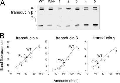
- Figure 5. Quantitative Western blotting of Pd-/- retinal extracts reveals decrease in transducin subunits expression. (A) Whole retinal extracts containing 100 fmol rhodopsin from WT and Pd-/- mice were separated on 10-20% polyacrylamide gels alongside 25, 50, 75, 100, 150 fmol purified transducin standard (1-5), and probed in Western blot analysis with antibodies against rod transducin subunits. (B) Fluorescence of the corresponding transducin standards bands from the top panel were plotted against the amounts of transducin in samples 1-5 (open circles), and fitted with sigmoid curves. Triangles represent the fluorescence of transducin bands from retina extracts. The number of determinations for each subunit is indicated in Table II .