 Explore
 Validate
 Learn
Learn Western blot
Antibody data
- Antibody Data
- Antigen structure
- References [1]
- Comments [0]
- Validations
- Western blot [3]
- Immunocytochemistry [1]
- Immunohistochemistry [9]
- Flow cytometry [3]
Submit
Validation data
Reference
Comment
Report error
- Product number
- TA502157 - Provider product page

- Provider
- OriGene
- Proper citation
- OriGene Cat#TA502157, RRID:AB_2622359
- Product name
- SAMHD1 mouse monoclonal antibody, clone OTI1A1 (formerly 1A1)
- Antibody type
- Monoclonal
- Description
- SAMHD1 mouse monoclonal antibody, clone OTI1A1 (formerly 1A1)
- Reactivity
- Canine
- Host
- Mouse
- Conjugate
- Unconjugated
- Epitope
- SAMHD1
- Isotype
- IgG
- Antibody clone number
- OTI1A1
- Vial size
- 100 µl
- Concentration
- 1.17 mg/ml
Submitted references Imaging HIV-1 Genomic DNA from Entry through Productive Infection.
Stultz RD, Cenker JJ, McDonald D
Journal of virology 2017 May 1;91(9)
Journal of virology 2017 May 1;91(9)
No comments: Submit comment
Supportive validation
- Submitted by
- OriGene (provider)
- Main image

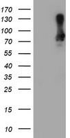
- Experimental details
- HEK293T cells were transfected with the pCMV6-ENTRY control (Left lane) or pCMV6-ENTRY SAMHD1 (RC206013, Right lane) cDNA for 48 hrs and lysed. Equivalent amounts of cell lysates (5 ug per lane) were separated by SDS-PAGE and immunoblotted with anti-SAMHD1.
- Validation comment
- WB
- Submitted by
- OriGene (provider)
- Main image
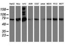
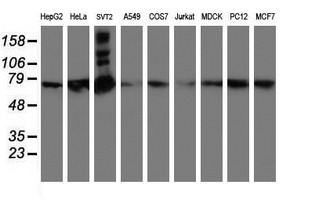
- Experimental details
- Western blot analysis of extracts (35ug) from 9 different cell lines by usin g anti-SAMHD1 monoclonal antibody (HepG2: human; HeLa: human; SVT2: mouse; A549: human; COS7: monkey; Jurkat: human; MDCK: canine; PC12: rat; MCF7: human).
- Validation comment
- WB
- Submitted by
- OriGene (provider)
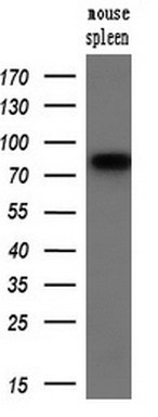
- Main image
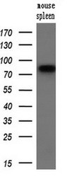
- Experimental details
- Western blot analysis of extracts (10ug) from a mouse tissue by using anti-SAMHD1 monoclonal antibody.(1:200)
- Validation comment
- WB
Supportive validation
- Submitted by
- OriGene (provider)
- Main image
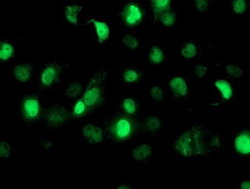
- Experimental details
- Anti-SAMHD1 mouse monoclonal antibody (TA502157) immunofluorescent staining of COS7 cells transiently transfected by pCMV6-ENTRY SAMHD1(RC206013).
- Validation comment
- IF

Supportive validation
- Submitted by
- OriGene (provider)
- Main image
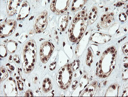
- Experimental details
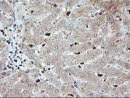
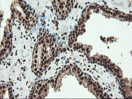
- Immunohistochemical staining of paraffin-embedded Human Kidney tissue within the normal limits using anti-SAMHD1 mouse monoclonal antibody. (Heat-induced epitope retrieval by 10mM citric buffer, pH6.0, 100C for 10min, TA502157)
- Validation comment
- IHC
- Submitted by
- OriGene (provider)
- Main image

- Experimental details
- Immunohistochemical staining of paraffin-embedded Human liver tissue within the normal limits using anti-SAMHD1 mouse monoclonal antibody. (Heat-induced epitope retrieval by 10mM citric buffer, pH6.0, 100C for 10min, TA502157)
- Validation comment
- IHC
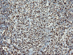
- Submitted by
- OriGene (provider)
- Main image
- Experimental details
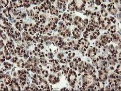
- Immunohistochemical staining of paraffin-embedded Carcinoma of Human thyroid tissue using anti-SAMHD1 mouse monoclonal antibody. (Heat-induced epitope retrieval by 10mM citric buffer, pH6.0, 100C for 10min, TA502157)
- Validation comment
- IHC

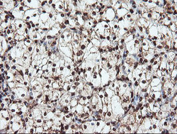
- Submitted by
- OriGene (provider)
- Main image
- Experimental details
- Immunohistochemical staining of paraffin-embedded Human pancreas tissue within the normal limits using anti-SAMHD1 mouse monoclonal antibody. (Heat-induced epitope retrieval by 10mM citric buffer, pH6.0, 100C for 10min, TA502157)
- Validation comment
- IHC

- Submitted by
- OriGene (provider)
- Main image

- Experimental details
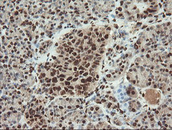
- Immunohistochemical staining of paraffin-embedded Adenocarcinoma of Human ovary tissue using anti-SAMHD1 mouse monoclonal antibody. (Heat-induced epitope retrieval by 10mM citric buffer, pH6.0, 100C for 10min, TA502157)
- Validation comment
- IHC
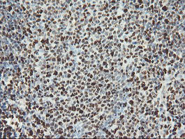
- Submitted by
- OriGene (provider)
- Main image
- Experimental details
- Immunohistochemical staining of paraffin-embedded Human tonsil within the normal limits using anti-SAMHD1 mouse monoclonal antibody. (Heat-induced epitope retrieval by 10mM citric buffer, pH6.0, 100C for 10min, TA502157)
- Validation comment
- IHC
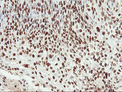
- Submitted by
- OriGene (provider)
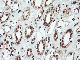
- Main image
- Experimental details
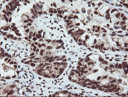
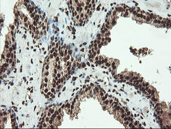
- Immunohistochemical staining of paraffin-embedded Carcinoma of Human kidney tissue using anti-SAMHD1 mouse monoclonal antibody. (Heat-induced epitope retrieval by 10mM citric buffer, pH6.0, 100C for 10min, TA502157)
- Validation comment
- IHC

- Submitted by
- OriGene (provider)
- Main image
- Experimental details
- Immunohistochemical staining of paraffin-embedded Carcinoma of Human bladder tissue using anti-SAMHD1 mouse monoclonal antibody. (Heat-induced epitope retrieval by 10mM citric buffer, pH6.0, 100C for 10min, TA502157)
- Validation comment
- IHC

- Submitted by
- OriGene (provider)
- Main image
- Experimental details
- Immunohistochemical staining of paraffin-embedded Carcinoma of Human prostate tissue using anti-SAMHD1 mouse monoclonal antibody. (Heat-induced epitope retrieval by 10mM citric buffer, pH6.0, 100C for 10min, TA502157)
- Validation comment
- IHC
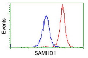
Supportive validation
- Submitted by
- OriGene (provider)
- Main image

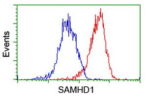
- Experimental details
- Flow cytometric Analysis of Hela cells, using anti-SAMHD1 antibody(TA502157),(Red), compared to a nonspecific negative control antibody,(Blue).
- Validation comment
- FC
- Submitted by
- OriGene (provider)
- Main image
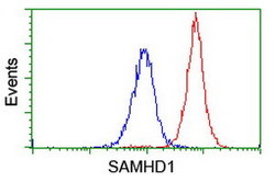
- Experimental details
- Flow cytometric Analysis of Jurkat cells, using anti-SAMHD1 antibody(TA502157),(Red), compared to a nonspecific negative control antibody,(Blue).
- Validation comment
- FC
- Submitted by
- OriGene (provider)
- Main image
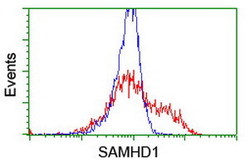
- Experimental details
- HEK293T cells transfected with either RC206013 overexpress plasmid(Red) or empty vector control plasmid(Blue) were immunostained by anti-SAMHD1 antibody(TA502157), and then analyzed by flow cytometry.
- Validation comment
- FC
