Explore
 Validate
 Learn
Learn Immunocytochemistry
Antibody data
- Antibody Data
- Antigen structure
- References [1]
- Comments [0]
- Validations
- Immunocytochemistry [1]
- Other assay [1]
Submit
Validation data
Reference
Comment
Report error
- Product number
- PA5-65793 - Provider product page

- Provider
- Invitrogen Antibodies
- Product name
- SEC24D Polyclonal Antibody
- Antibody type
- Polyclonal
- Antigen
- Recombinant protein fragment
- Description
- Immunogen sequence: IFLLANGLHMF LWLGVSSPPE LIQGIFNVPS FAHINTDMTL LPEVGNPYSQ QLRMIMGIIQ QKRPYSMKLT IVKQREQP Highest antigen sequence identity to the following orthologs - mouse 85%, rat 84%.
- Reactivity
- Human
- Host
- Rabbit
- Isotype
- IgG
- Vial size
- 100 μL
- Concentration
- 0.1 mg/mL
- Storage
- Store at 4°C short term. For long term storage, store at -20°C, avoiding freeze/thaw cycles.
Submitted references Cytisine is neuroprotective in female but not male 6-hydroxydopamine lesioned parkinsonian mice and acts in combination with 17-β-estradiol to inhibit apoptotic endoplasmic reticulum stress in dopaminergic neurons.
Zarate SM, Pandey G, Chilukuri S, Garcia JA, Cude B, Storey S, Salem NA, Bancroft EA, Hook M, Srinivasan R
Journal of neurochemistry 2021 May;157(3):710-726
Journal of neurochemistry 2021 May;157(3):710-726
No comments: Submit comment
Supportive validation
- Submitted by
- Invitrogen Antibodies (provider)
- Main image
- Experimental details
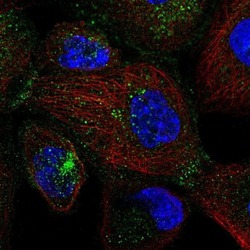
- Immunofluorescent staining of SEC24D in human cell line A-431 shows localization to vesicles. Samples were probed using a SEC24D Polyclonal Antibody (Product # PA5-65793).

Supportive validation
- Submitted by
- Invitrogen Antibodies (provider)
- Main image
- Experimental details
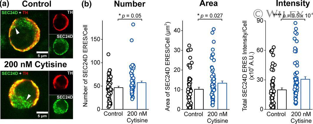
- 6 FIGURE Cytisine up-regulates Sec24D-containing ER exit sites (Sec24D-ERES) in mouse primary cultured dopaminergic (DA) neurons. (a) Representative confocal images of primary mouse DA neuron cell bodies stained for endogenous tyrosine hydroxylase (TH) (in red) and Sec24D-ERES (in green) are shown. Merged images for a control and 200 nM cytisine-treated DA neurons are shown along with separate images for TH and Sec24D staining. White arrowheads point to Sec24D-ERES puncta. Note the larger and more numerous Sec24D-ERES puncta following cytisine exposure. Scale bar is 5 mum. (b) Bar graphs quantifying the number, area, and intensity of Sec24D-ERES puncta in untreated (control) and cytisine-treated primary mouse DA cultures. All p values are based on Mann-Whitney test and error bars are S.E.M. ; n = 2 independent cultures and 60 DA neurons per condition; each independent culture is from embryonic day 14 embryos derived from multiple timed pregnant mice