Explore
 Validate
 Learn
Learn Western blot
Antibody data
- Antibody Data
- Antigen structure
- References [0]
- Comments [0]
- Validations
- Western blot [1]
- Immunohistochemistry [1]
Submit
Validation data
Reference
Comment
Report error
- Product number
- ACC-009-25UL - Provider product page

- Provider
- Invitrogen Antibodies
- Product name
- CaV3.3 (CACNA1I) Polyclonal Antibody
- Antibody type
- Polyclonal
- Antigen
- Other
- Reactivity
- Human, Mouse, Rat
- Host
- Rabbit
- Isotype
- IgG
- Vial size
- 25 µL
- Concentration
- 0.75 mg/mL
- Storage
- -20° C, Avoid Freeze/Thaw Cycles
No comments: Submit comment
Supportive validation
- Submitted by
- Invitrogen Antibodies (provider)
- Main image

- Experimental details
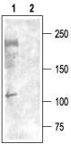
- Western blot analysisof rat brain membranes: - 1. Anti-CaV3.3 (CACNA1I) Antibody (#ACC-009), (1:200).2. Anti-CaV3.3 (CACNA1I) Antibody , preincubated with Cav3.3/CACNA1I Blocking Peptide (#BLP-CC009).
Supportive validation
- Submitted by
- Invitrogen Antibodies (provider)
- Main image
- Experimental details
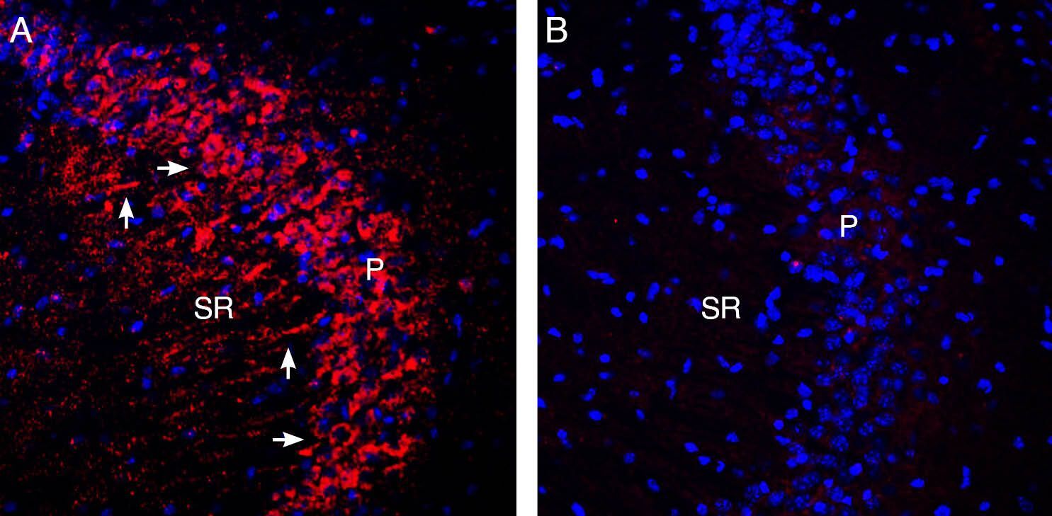
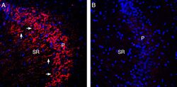
- Expression of CaV3.3 in mouse hippocampus. Immunohistochemical staining of perfusion-fixed frozen mouse brain sections with Anti-CaV3.3 (CACNA1I) Antibody (#ACC-009), (1:400), followed by donkey Anti-rabbit-Cy3. A. Staining in the hippocampal CA3 region, showed Cav3.3 immunoreactivity (red) appeared in soma of pyramidal neuron profiles (horizontal arrows) and in dendrites (vertical arrows) projecting into the stratum radiatum (SR). B. Pre-incubation of the Antibody with Cav3.3/CACNA1I Blocking Peptide (#BLP-CC009), suppressed staining. Cell nuclei are stained with DAPI (blue). P = pyramidal layer.