Explore
 Validate
 Learn
Learn Western blot
Antibody data
- Antibody Data
- Antigen structure
- References [2]
- Comments [0]
- Validations
- Western blot [1]
- Immunocytochemistry [2]
- Immunohistochemistry [5]
Submit
Validation data
Reference
Comment
Report error
- Product number
- GTX103193 - Provider product page

- Provider
- GeneTex
- Proper citation
- GeneTex Cat#GTX103193, RRID:AB_2037925
- Product name
- RRM2 antibody [N1C1]
- Antibody type
- Polyclonal
- Reactivity
- Human, Mouse, Rat
- Host
- Rabbit
Submitted references anti-EGFR capture mitigates EMT- and chemoresistance-associated heterogeneity in a resistance-profiling CTC platform.
The expression of ribonucleotide reductase M2 in the carcinogenesis of uterine cervix and its relationship with clinicopathological characteristics and prognosis of cancer patients.
Thege FI, Gruber CN, Cardle II, Cong SH, Lannin TB, Kirby BJ
Analytical biochemistry 2019 Feb 18;577:26-33
Analytical biochemistry 2019 Feb 18;577:26-33
The expression of ribonucleotide reductase M2 in the carcinogenesis of uterine cervix and its relationship with clinicopathological characteristics and prognosis of cancer patients.
Su YF, Wu TF, Ko JL, Tsai HT, Tee YT, Chien MH, Chou CH, Lin WL, Low HY, Chou MY, Yang SF, Wang PH
PloS one 2014;9(3):e91644
PloS one 2014;9(3):e91644
No comments: Submit comment
Supportive validation
- Submitted by
- GeneTex (provider)
- Main image

- Experimental details
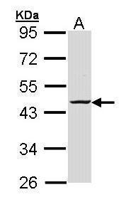
- Sample (30 ug of whole cell lysate) A: H1299 10% SDS PAGE GTX103193 diluted at 1:1000
- Validation comment
- WB
Supportive validation
- Submitted by
- GeneTex (provider)
- Main image
- Experimental details
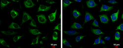
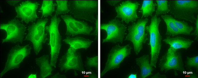
- RRM2 antibody [N1C1] detects RRM2 protein at cytoplasm by immunofluorescent analysis.Sample: HeLa cells were fixed in 4% paraformaldehyde at RT for 15 min.Green: RRM2 protein stained by RRM2 antibody [N1C1] (GTX103193) diluted at 1:500.Blue: Hoechst 33342 staining.Scale bar = 10 £gm.

- Submitted by
- GeneTex (provider)
- Main image
- Experimental details
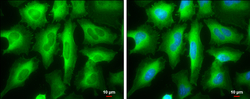
- RRM2 antibody [N1C1] detects RRM2 protein at cytoplasm by immunofluorescent analysis.Sample: HeLa cells were fixed in ice-cold MeOH for 5 min.Green: RRM2 protein stained by RRM2 antibody [N1C1] (GTX103193) diluted at 1:1000.Blue: Hoechst 33342 staining.Scale bar = 10 £gm.
Supportive validation
- Submitted by
- GeneTex (provider)
- Main image
- Experimental details
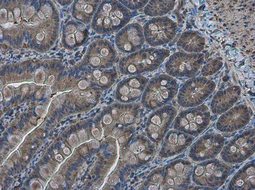
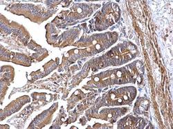
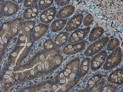
- RRM2 antibody [N1C1] detects RRM2 protein at cytoplasm on mouse intestine by immunohistochemical analysis. Sample: Paraffin-embedded mouse intestine. RRM2 antibody [N1C1] (GTX103193) diluted at 1:500.

- Submitted by
- GeneTex (provider)
- Main image
- Experimental details
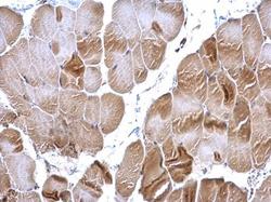
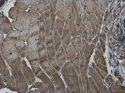
- RRM2 antibody [N1C1] detects RRM2 protein at cytoplasm on mouse muscle by immunohistochemical analysis. Sample: Paraffin-embedded mouse muscle. RRM2 antibody [N1C1] (GTX103193) diluted at 1:500.

- Submitted by
- GeneTex (provider)
- Main image
- Experimental details
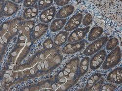
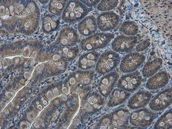
- RRM2 antibody [N1C1] detects RRM2 protein at cytoplasm in rat intestine by immunohistochemical analysis. Sample: Paraffin-embedded rat intestine. RRM2 antibody [N1C1] (GTX103193) diluted at 1:500.
- Submitted by
- GeneTex (provider)
- Main image
- Experimental details
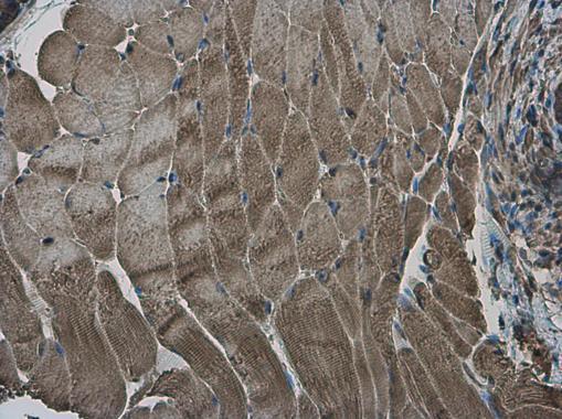
- RRM2 antibody [N1C1] detects RRM2 protein at cytoplasm in mouse prostate by immunohistochemical analysis. Sample: Paraffin-embedded mouse muscle. RRM2 antibody [N1C1] (GTX103193) diluted at 1:500.
- Submitted by
- GeneTex (provider)
- Main image
- Experimental details
- RRM2 antibody [N1C1] detects RRM2 protein at cytoplasm in rat intestine by immunohistochemical analysis. Sample: Paraffin-embedded rat intestine. RRM2 antibody [N1C1] (GTX103193) diluted at 1:500.