Explore
 Validate
 Learn
Learn Western blot
Antibody data
- Antibody Data
- Antigen structure
- References [3]
- Comments [0]
- Validations
- Western blot [1]
- ELISA [1]
- Immunocytochemistry [1]
- Immunoprecipitation [1]
- Immunohistochemistry [1]
Submit
Validation data
Reference
Comment
Report error
- Product number
- H00006241-M01 - Provider product page

- Provider
- Abnova Corporation
- Proper citation
- Abnova Corporation Cat#H00006241-M01, RRID:AB_509395
- Product name
- RRM2 monoclonal antibody (M01), clone 1E1
- Antibody type
- Monoclonal
- Description
- Mouse monoclonal antibody raised against a partial recombinant RRM2.
- Antigen sequence
MLSLRVPLAPITDPQQLQLSPLKGLSLVDKENTPP
ALSGTRVLASKTARRIFQEPTEPKTKAAAPGVEDE
PLLRENPRRFVIFPIEYHDIWQMYKKAEASFWTAE
EVDLS- Isotype
- IgG
- Antibody clone number
- 1E1
- Storage
- Store at -20°C or lower. Aliquot to avoid repeated freezing and thawing.
Submitted references RRM1 maintains centrosomal integrity via CHK1 and CDK1 signaling during replication stress.
Epstein-Barr virus episome stability is coupled to a delay in replication timing.
Quantitative phosphoproteome profiling of Wnt3a-mediated signaling network: indicating the involvement of ribonucleoside-diphosphate reductase M2 subunit phosphorylation at residue serine 20 in canonical Wnt signal transduction.
Kim SH, Park ER, Joo HY, Shen YN, Hong SH, Kim CH, Singh R, Lee KH, Shin HJ
Cancer letters 2014 May 1;346(2):249-56
Cancer letters 2014 May 1;346(2):249-56
Epstein-Barr virus episome stability is coupled to a delay in replication timing.
Zhou J, Snyder AR, Lieberman PM
Journal of virology 2009 Mar;83(5):2154-62
Journal of virology 2009 Mar;83(5):2154-62
Quantitative phosphoproteome profiling of Wnt3a-mediated signaling network: indicating the involvement of ribonucleoside-diphosphate reductase M2 subunit phosphorylation at residue serine 20 in canonical Wnt signal transduction.
Tang LY, Deng N, Wang LS, Dai J, Wang ZL, Jiang XS, Li SJ, Li L, Sheng QH, Wu DQ, Li L, Zeng R
Molecular & cellular proteomics : MCP 2007 Nov;6(11):1952-67
Molecular & cellular proteomics : MCP 2007 Nov;6(11):1952-67
No comments: Submit comment
Supportive validation
- Submitted by
- Abnova Corporation (provider)
- Main image

- Experimental details
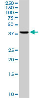
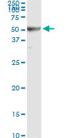
- RRM2 monoclonal antibody (M01), clone 1E1. Western Blot analysis of RRM2 expression in HepG2 ( Cat # L019V1 ).
Supportive validation
- Submitted by
- Abnova Corporation (provider)
- Main image
- Experimental details
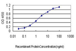
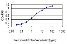
- Detection limit for recombinant GST tagged RRM2 is approximately 0.03ng/ml as a capture antibody.
- Validation comment
- Sandwich ELISA (Recombinant protein)
- Protocol
- Protocol
Supportive validation
- Submitted by
- Abnova Corporation (provider)
- Main image
- Experimental details
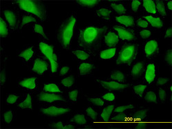
- Immunofluorescence of monoclonal antibody to RRM2 on HeLa cell. [antibody concentration 10 ug/ml]
- Validation comment
- Immunofluorescence
- Protocol
- Protocol

Supportive validation
- Submitted by
- Abnova Corporation (provider)
- Main image

- Experimental details
- Immunoprecipitation of RRM2 transfected lysate using anti-RRM2 monoclonal antibody and Protein A Magnetic Bead (U0007), and immunoblotted with RRM2 MaxPab rabbit polyclonal antibody.
- Validation comment
- Immunoprecipitation
- Protocol
- Protocol
Supportive validation
- Submitted by
- Abnova Corporation (provider)
- Main image
- Experimental details
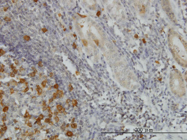
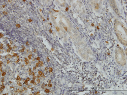
- Immunoperoxidase of monoclonal antibody to RRM2 on formalin-fixed paraffin-embedded human stomach. [antibody concentration 1.5 ug/ml]
- Validation comment
- Immunohistochemistry (Formalin/PFA-fixed paraffin-embedded sections)
- Protocol
- Protocol