 Explore
 Validate
 Learn
Learn Western blot
Antibody data
- Antibody Data
- Antigen structure
- References [0]
- Comments [0]
- Validations
- Western blot [3]
- Immunocytochemistry [3]
- Immunohistochemistry [4]
Submit
Validation data
Reference
Comment
Report error
- Product number
- PA5-27856 - Provider product page

- Provider
- Invitrogen Antibodies
- Product name
- RRM2 Polyclonal Antibody
- Antibody type
- Polyclonal
- Antigen
- Recombinant protein fragment
- Description
- Predicted reactivity: Mouse (89%), Rat (88%), Zebrafish (86%), Bovine (94%).
- Concentration
- 1.18 mg/mL
No comments: Submit comment
Supportive validation
- Submitted by
- Invitrogen Antibodies (provider)
- Main image

- Experimental details
- Western blot analysis of RRM2 using 30 µg of H1299 lysate. Samples were loaded onto a 10% SDS-PAGE gel and probed with a RRM2 polyclonal antibody (Product # PA5-27856) at a dilution of 1:1000.

- Submitted by
- Invitrogen Antibodies (provider)
- Main image

- Experimental details
- Knockdown of RRM2 was achieved by transfecting A549 cells with RRM2 specific siRNAs (Silencer® select Product # s12360, s12361). Western blot analysis (Fig. a) was performed using whole cell extracts from the RRM2 knockdown cells (lane 3), non-specific scrambled siRNA transfected cells (lane 2) and un-transfected cells (lane 1). The blots were probed with RRM2 Polyclonal Antibody (Product # PA5-27856, 1µg/ml) and Goat anti-Rabbit IgG (H+L) Superclonal™ Secondary Antibody, HRP conjugate (Product # A27036, 0.25µg/ml, 1:4000 dilution). Densitometry analysis of this western blot is shown in histogram (Fig b). Decrease in signal upon siRNA mediated knock down confirms that antibody is specific to RRM2.

- Submitted by
- Invitrogen Antibodies (provider)
- Main image
- Experimental details
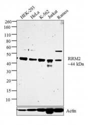
- Western blot analysis was performed on whole cell extracts (30 µg lysate) of HEK-293 (Lane 1), HeLa (Lane 2), K-562 (Lane 3), Jurkat (Lane 4) and Ramos (Lane 5). The blot was probed with Anti-RRM2 Polyclonal Antibody (Product # PA5-27856, 1µg/ml) and detected by chemiluminescence using Goat anti-Rabbit IgG (H+L) Superclonal™ Secondary Antibody, HRP conjugate (Product # A27036, 0.25 µg/ml, 1:4000 dilution). A 44 kDa band corresponding to RRM2 was observed across all the cell lines tested.

Supportive validation
- Submitted by
- Invitrogen Antibodies (provider)
- Main image

- Experimental details
- Immunocytochemistry-Immunofluorescence analysis of RRM2 was performed in HeLa cells fixed in 4% paraformaldehyde at RT for 15 min. Green: RRM2 Polyclonal Antibody (Product # PA5-27856) diluted at 1:500. Blue: Hoechst 33342 staining. Scale bar = 10 µm.

- Submitted by
- Invitrogen Antibodies (provider)
- Main image

- Experimental details
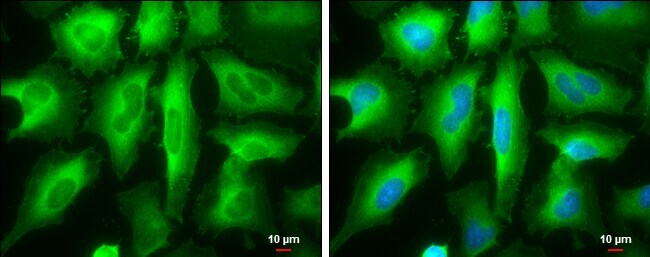
- Immunocytochemistry-Immunofluorescence analysis of RRM2 was performed in HeLa cells fixed in ice-cold MeOH for 5 min. Green: RRM2 Polyclonal Antibody (Product # PA5-27856) diluted at 1:1000. Blue: Hoechst 33342 staining. Scale bar = 10 µm.
- Submitted by
- Invitrogen Antibodies (provider)
- Main image

- Experimental details
- Immunofluorescence analysis of RRM2 was performed using 70% confluent log phase A549 cells. The cells were fixed with 4% paraformaldehyde for 10 minutes, permeabilized with 0.1% Triton™ X-100 for 15 minutes, and blocked with 1% BSA for 1 hour at room temperature. The cells were labeled with RRM2 Polyclonal Antibody (Product # PA5-27856) at 5 µg/mL in 0.1% BSA, incubated at 4 degree celsius overnight and then labeled with Goat anti-Rabbit IgG (H+L) Superclonal™ Secondary Antibody, Alexa Fluor® 488 conjugate (Product # A27034) at a dilution of 1:2000 for 45 minutes at room temperature (Panel a: green).Nuclei (Panel b: blue) were stained with SlowFade® Gold Antifade Mountant with DAPI (Product # S36938). F-actin (Panel c: red) was stained with Rhodamine Phalloidin (Product # R415, 1:300). Panel d represents the merged image showing predominant cytosolic localization. Panel e represents control cells with no primary antibody to assess background. The images were captured at 60X magnification.

Supportive validation
- Submitted by
- Invitrogen Antibodies (provider)
- Main image

- Experimental details
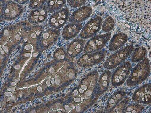
- Immunohistochemistry (Paraffin) analysis of RRM2 was performed in paraffin-embedded rat intestine tissue using RRM2 Polyclonal Antibody (Product # PA5-27856) at a dilution of 1:500.
- Submitted by
- Invitrogen Antibodies (provider)
- Main image

- Experimental details
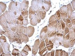
- Immunohistochemistry (Paraffin) analysis of RRM2 was performed in paraffin-embedded mouse muscle tissue using RRM2 Polyclonal Antibody (Product # PA5-27856) at a dilution of 1:500.

- Submitted by
- Invitrogen Antibodies (provider)
- Main image
- Experimental details
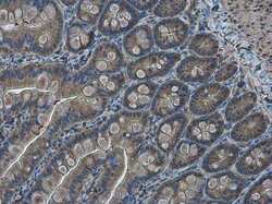
- Immunohistochemistry (Paraffin) analysis of RRM2 was performed in paraffin-embedded rat intestine tissue using RRM2 Polyclonal Antibody (Product # PA5-27856) at a dilution of 1:500.

- Submitted by
- Invitrogen Antibodies (provider)
- Main image
- Experimental details
- RRM2 Polyclonal Antibody detects RRM2 protein at cytoplasm on mouse muscle by immunohistochemical analysis. Sample: Paraffin-embedded mouse muscle. RRM2 Polyclonal Antibody (Product # PA5-27856) diluted at 1:500. Antigen Retrieval: EDTA based buffer, pH 8.0, 15 min.
