Explore
 Validate
 Learn
Learn Western blot
 Immunocytochemistry
Antibody data
- Antibody Data
- Antigen structure
- References [0]
- Comments [0]
- Validations
- Immunocytochemistry [1]
- Immunohistochemistry [7]
- Flow cytometry [5]
Submit
Validation data
Reference
Comment
Report error
- Product number
- MA5-49271 - Provider product page

- Provider
- Invitrogen Antibodies
- Product name
- eRF1 Monoclonal Antibody (3B6)
- Antibody type
- Monoclonal
- Antigen
- Recombinant full-length protein
- Description
- Adding 0.2 mL of distilled water will yield a concentration of 500 µg/mL. Positive Control - WB: human Hela whole cell, human Jurkat whole cell, human K562 whole cell, human Raji whole cell, human HEPG2 whole cell, rat PC-12 whole cell, mouse RAW2647 whole cell. IHC: human tonsil tissue, human ovarian cancer tissue, human breast cancer tissue, human breast cancer tissue, human liver cancer tissue, human lung cancer tissue, rat kidney tissue. ICC/IF: MCF-7 cell. Flow: HEPA1-6 cell, CACO-2 cell, RH35 cell.|Store at -20°C for one year from date of receipt. After reconstitution, at 4°C for one month. It can also be aliquotted and stored frozen at -20°C for six months. Avoid repeated freeze-thaw cycles.
- Reactivity
- Human, Mouse, Rat
- Host
- Mouse
- Isotype
- IgG
- Antibody clone number
- 3B6
- Vial size
- 100 μg
- Concentration
- 500 μg/mL
- Storage
- -20°C
No comments: Submit comment
Supportive validation
- Submitted by
- Invitrogen Antibodies (provider)
- Main image

- Experimental details
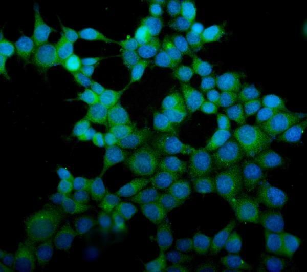
- Immunocytochemistry analysis of eRF1 in MCF-7 cells. Enzyme antigen retrieval was performed using IHC enzyme antigen retrieval reagent for 15 mins. The cells were blocked with 10% goat serum. Samples were then incubated in eRF1 Monoclonal antibody (Product # MA5-49271) using a dilution of 5 μg/mL. DyLight®488 Conjugated Goat Anti-Mouse IgG was used as secondary antibody at 1:100 dilution and incubated for 30 minutes at 37°C. The section was counterstained with DAPI. Visualize using a fluorescence microscope and filter sets appropriate for the label used.
Supportive validation
- Submitted by
- Invitrogen Antibodies (provider)
- Main image
- Experimental details
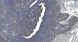
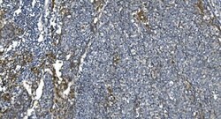
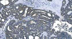
- Immunohistochemistry analysis of eRF1 in paraffin-embedded section of human tonsil tissue. Heat mediated antigen retrieval was performed in EDTA buffer (pH 8.0, epitope retrieval solution). The tissue section was blocked with 10% goat serum. Samples were incubated with eRF1 Monoclonal antibody (Product # MA5-49271) using a dilution of 2 μg/mL overnight at 4°C. Biotinylated goat anti-mouse IgG was used as secondary antibody and incubated for 30 minutes at 37°C. The tissue section was developed using Strepavidin-Biotin-Complex (SABC) with DAB as the chromogen.
- Submitted by
- Invitrogen Antibodies (provider)
- Main image
- Experimental details
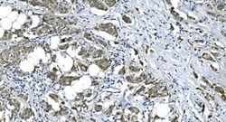
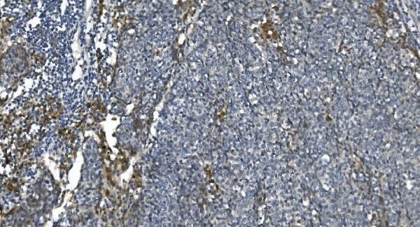
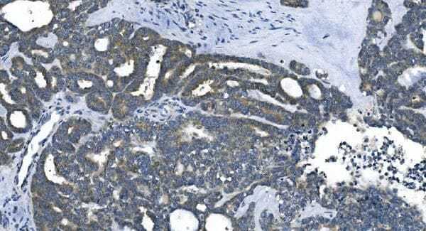
- Immunohistochemistry analysis of eRF1 in paraffin-embedded section of human breast cancer tissue. Heat mediated antigen retrieval was performed in EDTA buffer (pH 8.0, epitope retrieval solution). The tissue section was blocked with 10% goat serum. Samples were incubated with eRF1 Monoclonal antibody (Product # MA5-49271) using a dilution of 2 μg/mL overnight at 4°C. Biotinylated goat anti-mouse IgG was used as secondary antibody and incubated for 30 minutes at 37°C. The tissue section was developed using Strepavidin-Biotin-Complex (SABC) with DAB as the chromogen.
- Submitted by
- Invitrogen Antibodies (provider)
- Main image
- Experimental details
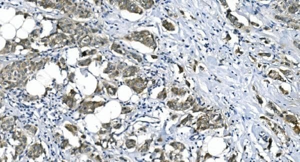
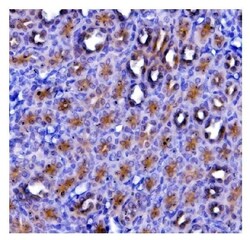
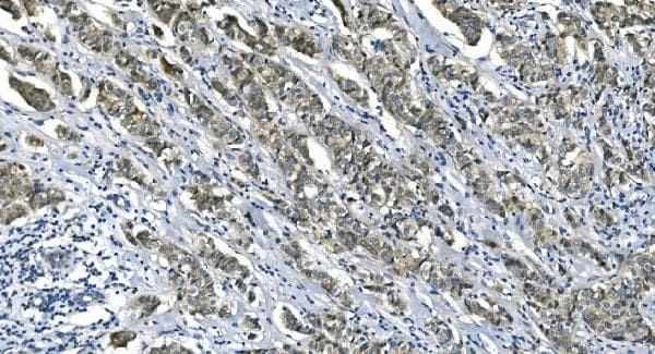
- Immunohistochemistry analysis of eRF1 in paraffin-embedded section of human liver cancer tissue. Heat mediated antigen retrieval was performed in EDTA buffer (pH 8.0, epitope retrieval solution). The tissue section was blocked with 10% goat serum. Samples were incubated with eRF1 Monoclonal antibody (Product # MA5-49271) using a dilution of 2 μg/mL overnight at 4°C. Biotinylated goat anti-mouse IgG was used as secondary antibody and incubated for 30 minutes at 37°C. The tissue section was developed using Strepavidin-Biotin-Complex (SABC) with DAB as the chromogen.
- Submitted by
- Invitrogen Antibodies (provider)
- Main image
- Experimental details
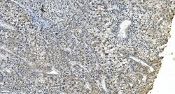
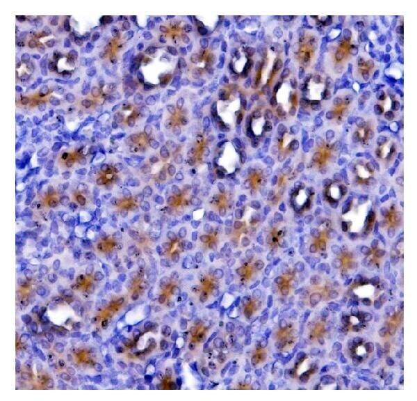
- Immunohistochemistry analysis of eRF1 in paraffin-embedded section of human lung cancer tissue. Heat mediated antigen retrieval was performed in EDTA buffer (pH 8.0, epitope retrieval solution). The tissue section was blocked with 10% goat serum. Samples were incubated with eRF1 Monoclonal antibody (Product # MA5-49271) using a dilution of 2 μg/mL overnight at 4°C. Biotinylated goat anti-mouse IgG was used as secondary antibody and incubated for 30 minutes at 37°C. The tissue section was developed using Strepavidin-Biotin-Complex (SABC) with DAB as the chromogen.
- Submitted by
- Invitrogen Antibodies (provider)
- Main image
- Experimental details
- Immunohistochemistry analysis of eRF1 in paraffin-embedded section of rat kidney tissue. Heat mediated antigen retrieval was performed in EDTA buffer (pH 8.0, epitope retrieval solution). The tissue section was blocked with 10% goat serum. Samples were incubated with eRF1 Monoclonal antibody (Product # MA5-49271) using a dilution of 2 μg/mL overnight at 4°C. Biotinylated goat anti-mouse IgG was used as secondary antibody and incubated for 30 minutes at 37°C. The tissue section was developed using Strepavidin-Biotin-Complex (SABC) with DAB as the chromogen.
- Submitted by
- Invitrogen Antibodies (provider)
- Main image
- Experimental details
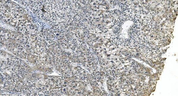
- Immunohistochemistry analysis of eRF1 in paraffin-embedded section of human ovarian cancer tissue. Heat mediated antigen retrieval was performed in EDTA buffer (pH 8.0, epitope retrieval solution). The tissue section was blocked with 10% goat serum. Samples were incubated with eRF1 Monoclonal antibody (Product # MA5-49271) using a dilution of 2 μg/mL overnight at 4°C. Biotinylated goat anti-mouse IgG was used as secondary antibody and incubated for 30 minutes at 37°C. The tissue section was developed using Strepavidin-Biotin-Complex (SABC) with DAB as the chromogen.
- Submitted by
- Invitrogen Antibodies (provider)
- Main image

- Experimental details
- Immunohistochemistry analysis of eRF1 in paraffin-embedded section of human breast cancer tissue. Heat mediated antigen retrieval was performed in EDTA buffer (pH 8.0, epitope retrieval solution). The tissue section was blocked with 10% goat serum. Samples were incubated with eRF1 Monoclonal antibody (Product # MA5-49271) using a dilution of 2 μg/mL overnight at 4°C. Biotinylated goat anti-mouse IgG was used as secondary antibody and incubated for 30 minutes at 37°C. The tissue section was developed using Strepavidin-Biotin-Complex (SABC) with DAB as the chromogen.
Supportive validation
- Submitted by
- Invitrogen Antibodies (provider)
- Main image
- Experimental details
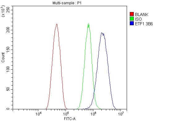
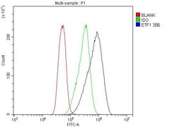
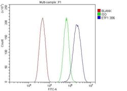
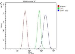
- Flow cytometry analysis of eRF1 in CACO-2 cells using eRF1 Monoclonal Antibody (3B6) (Product # MA5-49271), shown in overlay histogram (blue line). To facilitate intracellular staining, cells were fixed with 4% paraformaldehyde and permeabilized with permeabilization buffer. The cells were blocked with 10% normal goat serum, and incubated with the primary antibody (1 μg/1x10^6 cells) for 30 min at 20°C. DyLight 488 conjugated goat anti-mouse IgG (5-10 µg/1x10^6 cells) was used as secondary antibody for 30 minutes at 20°C. Isotype control antibody (Green line) was mouse IgG (1 µg/1x10^6) used under the same conditions. Unlabelled sample without incubation with primary antibody and secondary antibody (Red line) was used as a blank control.

- Submitted by
- Invitrogen Antibodies (provider)
- Main image
- Experimental details
- Flow cytometry of eRF1 in HEPA1-6 cells (Blue line). The cells were blocked with 10% normal goat serum, and then incubated with eRF1 monoclonal antibody (Product # MA5-49271) (1 µg/1x10^6 cells) for 30 min at 20°C. Isotype control antibody (Green line) was mouse IgG (1 μg/1x10^6 cells) used under the same conditions. Unlabeled sample (Red line) was also used as a control. DyLight®488 conjugated goat anti-mouse IgG (5-10 μg/1x10^6 cells) was used as secondary antibody for 30 minutes at 20°C.
- Submitted by
- Invitrogen Antibodies (provider)
- Main image
- Experimental details
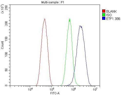
- Flow cytometry of eRF1 in RH35 cells (Blue line). The cells were blocked with 10% normal goat serum, and then incubated with eRF1 monoclonal antibody (Product # MA5-49271) (1 µg/1x10^6 cells) for 30 min at 20°C. Isotype control antibody (Green line) was mouse IgG (1 µg/1x10^6 cells) used under the same conditions. Unlabeled sample (Red line) was also used as a control. DyLight®488 conjugated goat anti-mouse IgG (5-10 μg/1x10^6 cells) was used as secondary antibody for 30 minutes at 20°C.
- Submitted by
- Invitrogen Antibodies (provider)
- Main image

- Experimental details
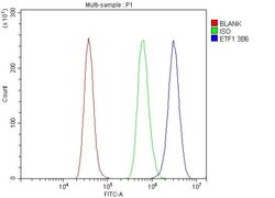
- Flow cytometry analysis of eRF1 in RH35 cells using eRF1 Monoclonal Antibody (3B6) (Product # MA5-49271), shown in overlay histogram (blue line). To facilitate intracellular staining, cells were fixed with 4% paraformaldehyde and permeabilized with permeabilization buffer. The cells were blocked with 10% normal goat serum, and incubated with the primary antibody (1 μg/1x10^6 cells) for 30 min at 20°C. DyLight 488 conjugated goat anti-mouse IgG (5-10 µg/1x10^6 cells) was used as secondary antibody for 30 minutes at 20°C. Isotype control antibody (Green line) was mouse IgG (1 µg/1x10^6) used under the same conditions. Unlabelled sample without incubation with primary antibody and secondary antibody (Red line) was used as a blank control.

- Submitted by
- Invitrogen Antibodies (provider)
- Main image
- Experimental details
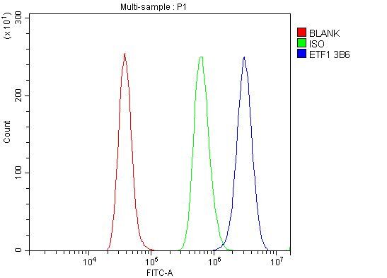
- Flow cytometry analysis of eRF1 in HEPA1-6 cells using eRF1 Monoclonal Antibody (3B6) (Product # MA5-49271), shown in overlay histogram (blue line). To facilitate intracellular staining, cells were fixed with 4% paraformaldehyde and permeabilized with permeabilization buffer. The cells were blocked with 10% normal goat serum, and incubated with the primary antibody (1 μg/1x10^6 cells) for 30 min at 20°C. DyLight 488 conjugated goat anti-mouse IgG (5-10 µg/1x10^6 cells) was used as secondary antibody for 30 minutes at 20°C. Isotype control antibody (Green line) was mouse IgG (1 µg/1x10^6) used under the same conditions. Unlabelled sample without incubation with primary antibody and secondary antibody (Red line) was used as a blank control.