 Explore
 Validate
 Learn
Learn Western blot
 ELISA
ELISA Immunocytochemistry
Antibody data
- Antibody Data
- Antigen structure
- References [0]
- Comments [0]
- Validations
- Western blot [3]
- ELISA [3]
- Immunohistochemistry [6]
Submit
Validation data
Reference
Comment
Report error
- Product number
- LS-C682371 - Provider product page

- Provider
- LSBio
- Product name
- RALA / RAL Antibody (clone 4G8C7) LS-C682371
- Antibody type
- Monoclonal
- Description
- Purified
- Reactivity
- Human
- Host
- Mouse
- Isotype
- IgG
- Antibody clone number
- 4G8C7
- Storage
- Short term: store at 4°C. Long term: store at -20°C.
No comments: Submit comment
Enhanced validation
- Submitted by
- LSBio (provider)
- Enhanced method
- Genetic validation
- Main image

- Experimental details
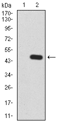
- Western blot analysis using RALA mAb against HEK293 (1) and RALA (AA: 71-203)-hIgGFc transfected HEK293 (2) cell lysate.
- Submitted by
- LSBio (provider)
- Enhanced method
- Genetic validation
- Main image

- Experimental details
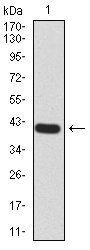
- Western blot analysis using RALA mAb against human RALA (AA: 71-203) recombinant protein. (Expected MW is 41.5 kDa)
- Submitted by
- LSBio (provider)
- Enhanced method
- Genetic validation
- Main image
- Experimental details
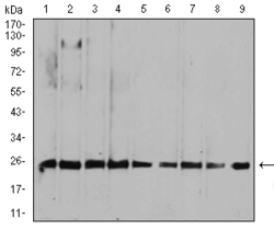
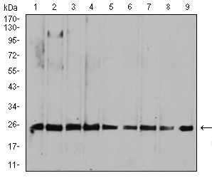
- Western blot analysis using RALA mouse mAb against HepG2 (1), MCF-7 (2), A549 (3), K562 (4), Raji (5), MOLT4 (6), Hela (7), COS7 (8), and NIH3T3 (9) cell lysate.
Supportive validation
- Submitted by
- LSBio (provider)
- Enhanced method
- Genetic validation
- Main image
- Experimental details
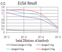
- Black line: Control Antigen (100 ng);Purple line: Antigen(10ng);Blue line: Antigen (50 ng);Red line: Antigen (100 ng);
- Submitted by
- LSBio (provider)
- Main image
- Experimental details
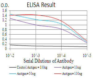
- Black line: Control Antigen (100 ng);Purple line: Antigen(10ng);Blue line: Antigen (50 ng);Red line: Antigen (100 ng);
- Submitted by
- LSBio (provider)
- Main image

- Experimental details
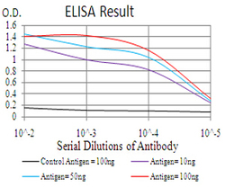
- Black line: Control Antigen (100 ng);Purple line: Antigen(10ng);Blue line: Antigen (50 ng);Red line: Antigen (100 ng);

Supportive validation
- Submitted by
- LSBio (provider)
- Enhanced method
- Genetic validation
- Main image

- Experimental details
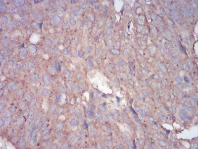
- Immunohistochemical analysis of paraffin-embedded ovarian cancer tissues using RALA mouse mAb with DAB staining.
- Submitted by
- LSBio (provider)
- Enhanced method
- Genetic validation
- Main image
- Experimental details
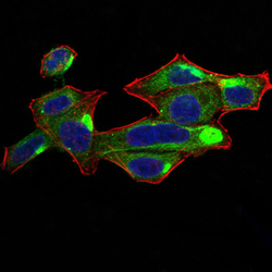
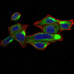
- Immunofluorescence analysis of Hela cells using RALA mouse mAb (green). Blue: DRAQ5 fluorescent DNA dye. Red: Actin filaments have been labeled with Alexa Fluor- 555 phalloidin. Secondary antibody from Fisher

- Submitted by
- LSBio (provider)
- Enhanced method
- Genetic validation
- Main image

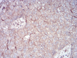
- Experimental details
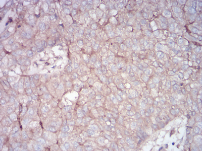
- Immunohistochemical analysis of paraffin-embedded bladder cancer tissues using RALA mouse mAb with DAB staining.
- Submitted by
- LSBio (provider)
- Enhanced method
- Genetic validation
- Main image

- Experimental details
- Immunohistochemical analysis of paraffin-embedded ovarian cancer tissues using RALA mouse mAb with DAB staining.

- Submitted by
- LSBio (provider)
- Enhanced method
- Genetic validation
- Main image

- Experimental details
- Immunofluorescence analysis of Hela cells using RALA mouse mAb (green). Blue: DRAQ5 fluorescent DNA dye. Red: Actin filaments have been labeled with Alexa Fluor- 555 phalloidin. Secondary antibody from Fisher
- Submitted by
- LSBio (provider)
- Enhanced method
- Genetic validation
- Main image
- Experimental details
- Immunohistochemical analysis of paraffin-embedded bladder cancer tissues using RALA mouse mAb with DAB staining.
