Explore
 Validate
 Learn
Learn Western blot
Antibody data
- Antibody Data
- Antigen structure
- References [1]
- Comments [0]
- Validations
- Western blot [1]
- Immunohistochemistry [1]
- Other assay [3]
Submit
Validation data
Reference
Comment
Report error
- Product number
- PA5-106907 - Provider product page

- Provider
- Invitrogen Antibodies
- Product name
- EXT1 Polyclonal Antibody
- Antibody type
- Polyclonal
- Antigen
- Synthetic peptide
- Description
- Antibody detects endogenous levels of total EXT1.
- Reactivity
- Human, Mouse, Rat
- Host
- Rabbit
- Isotype
- IgG
- Vial size
- 100 μL
- Concentration
- 1 mg/mL
- Storage
- -20°C
Submitted references Prevalence of neural epidermal growth factor-like 1- and exostosin 1/exostosin 2-associated membranous nephropathy: a single-center retrospective study in Japan.
Iwakura T, Ema C, Isobe S, Fujikura T, Ohashi N, Kato A, Yasuda H
Scientific reports 2022 Feb 22;12(1):2967
Scientific reports 2022 Feb 22;12(1):2967
No comments: Submit comment
Supportive validation
- Submitted by
- Invitrogen Antibodies (provider)
- Main image

- Experimental details
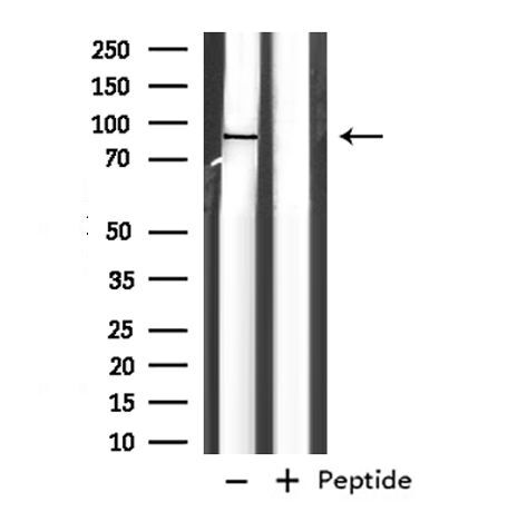
- Western blot analysis of EXT1 in rat brain. Samples were incubated with EXT1 polyclonal antibody (Product # PA5-106907).
Supportive validation
- Submitted by
- Invitrogen Antibodies (provider)
- Main image
- Experimental details
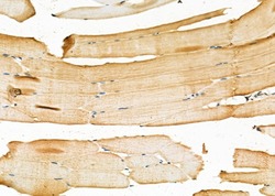
- Immunohistochemistry analysis of EXT1 in mouse muscle tissue. The sample was formaldehyde fixed and a heat mediated antigen retrieval step in citrate buffer was performed. Samples were incubated with EXT1 polyclonal antibody (Product # PA5-106907) using a dilution of 1:100 (4°C overnight) followed by HRP conjugated anti-Rabbit secondary antibody.

Supportive validation
- Submitted by
- Invitrogen Antibodies (provider)
- Main image
- Experimental details
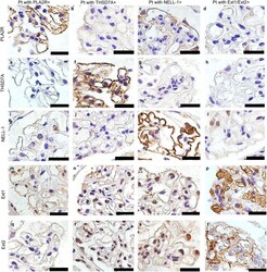
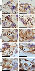
- Figure 3 Immunohistochemical analysis for Phospholipase A2 receptor (PLA2R), Thrombospondin type-1 domain-containing 7A (THSD7A), Neural epidermal growth factor-like 1 protein (NELL-1), Exostosin (Ext) 1 and Ext2. Representative cases of immunostaining of PLA2R ( a - d ), THSD7A ( e - h ), NELL-1 ( i - l ), Ext1 ( m - p ) and Ext2 ( q - t ) in patients with primary membranous nephropathy. Original magnification: x1000. Scar bar 25 mum. Pt patient.

- Submitted by
- Invitrogen Antibodies (provider)
- Main image
- Experimental details
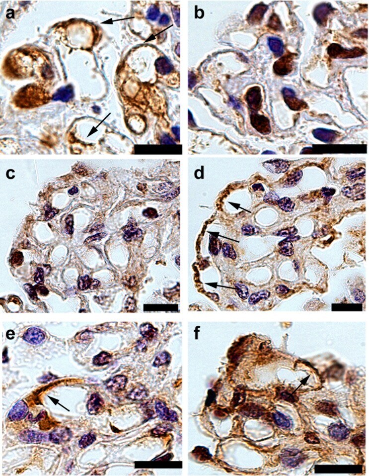
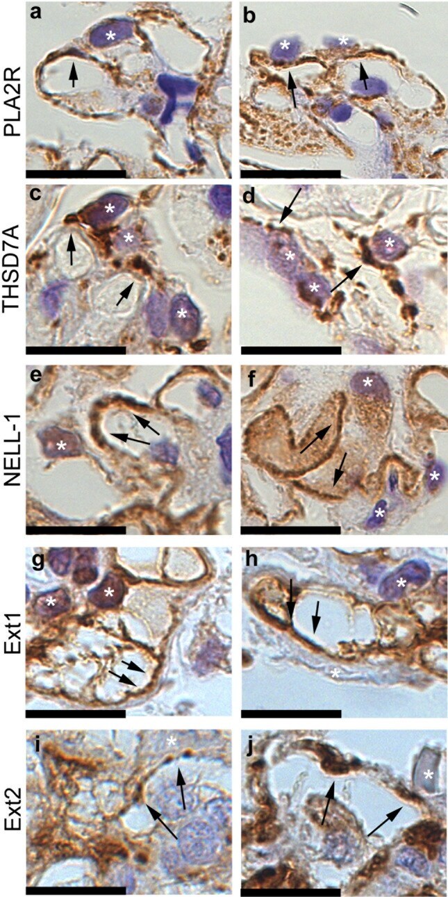
- Figure 4 Immunohistochemical analysis for case of Phospholipase A2 receptor (PLA2R)-, Thrombospondin type-1 domain-containing 7A (THSD7A)-, Neural epidermal growth factor-like 1 protein (NELL-1)-, Exostosin (Ext) 1- and Ext2-associated membranous nephropathy with high magnification. Photographs of immunostaining of PLA2R ( a , b ), THSD7A ( c , d ), NELL-1 ( e , f ), Ext1( g , h ) and Ext2 ( i , j ) with high magnification. The localization of enhanced antigens was shown with arrows. Asterisks; podocytes. Scar bar 12.5 mum.
- Submitted by
- Invitrogen Antibodies (provider)
- Main image
- Experimental details
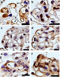
- Figure 6 Immunohistochemical analysis of exostosin (Ext) 1 and Ext2 in the atypical cases of Ext1/Ext2-associated membranous nephropathy (MN). Photographs of immunostaining of Ext1 ( a , c , e ) and Ext2 ( b , d , f ). A sample in primary MN showed positive staining of Ext1 ( a ), but negative staining of Ext2 ( b ). In lupus MN patients, a sample showed negative staining of Ext1 ( c ), but segmental, positive staining of Ext2 ( d ), while another sample showed segmental, positive staining of Ext1 ( e ) and Ext2 ( f ). Original magnification: x1000. Scar bar 12.5 mum.