Explore
 Validate
 Learn
Learn Western blot
Antibody data
- Antibody Data
- Antigen structure
- References [0]
- Comments [0]
- Validations
- Western blot [1]
- Immunohistochemistry [10]
Submit
Validation data
Reference
Comment
Report error
- Product number
- LS-C782440 - Provider product page

- Provider
- LSBio
- Product name
- SPI1 / PU.1 Antibody (clone PU1/2146) LS-C782440
- Antibody type
- Monoclonal
- Description
- Protein G affinity chromatography
- Reactivity
- Human
- Host
- Mouse
- Isotype
- IgG
- Antibody clone number
- PU1/2146
- Storage
- Store at 2-8°C.
No comments: Submit comment
Enhanced validation
- Submitted by
- LSBio (provider)
- Enhanced method
- Genetic validation
- Main image
- Experimental details
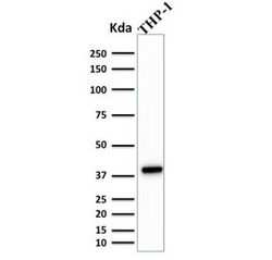
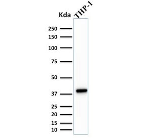
- Western blot testing of human THP-1 cell lysate with PU.1 antibody (clone PU1/2146). Predicted molecular weight ~31 kDa but routinely observed at ~40 kDa.
Enhanced validation
- Submitted by
- LSBio (provider)
- Enhanced method
- Genetic validation
- Main image
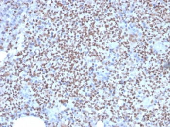
- Experimental details
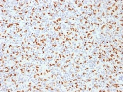
- IHC staining of FFPE human lymph node with SOX9 antibody (clone PU1/2146). HIER: boil tissue sections in pH6, 10mM citrate buffer, for 10-20 min followed by cooling at RT for 20 min.
- Submitted by
- LSBio (provider)
- Enhanced method
- Genetic validation
- Main image
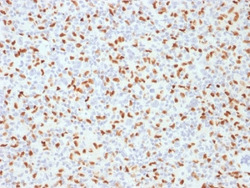
- Experimental details
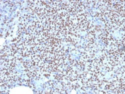
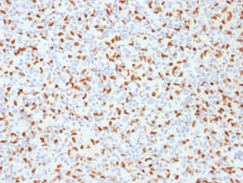
- IHC staining of FFPE Hodgkins lymphoma with SOX9 antibody (clone PU1/2146). HIER: boil tissue sections in pH6, 10mM citrate buffer, for 10-20 min followed by cooling at RT for 20 min.

- Submitted by
- LSBio (provider)
- Enhanced method
- Genetic validation
- Main image

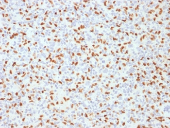
- Experimental details
- IHC staining of FFPE human lymph node with SOX9 antibody (clone PU1/2146). HIER: boil tissue sections in pH6, 10mM citrate buffer, for 10-20 min followed by cooling at RT for 20 min.

- Submitted by
- LSBio (provider)
- Enhanced method
- Genetic validation
- Main image

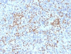
- Experimental details
- IHC staining of FFPE Hodgkins lymphoma with SOX9 antibody (clone PU1/2146). HIER: boil tissue sections in pH6, 10mM citrate buffer, for 10-20 min followed by cooling at RT for 20 min.
- Submitted by
- LSBio (provider)
- Enhanced method
- Genetic validation
- Main image

- Experimental details
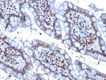
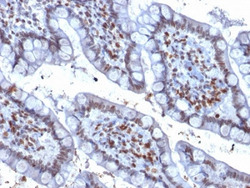
- IHC staining of FFPE human colon carcinoma with SOX9 antibody (clone PU1/2146). HIER: boil tissue sections in pH6, 10mM citrate buffer, for 10-20 min followed by cooling at RT for 20 min.

- Submitted by
- LSBio (provider)
- Enhanced method
- Genetic validation
- Main image

- Experimental details
- IHC staining of FFPE human spleen with SOX9 antibody (clone PU1/2146). HIER: boil tissue sections in pH6, 10mM citrate buffer, for 10-20 min followed by cooling at RT for 20 min.

- Submitted by
- LSBio (provider)
- Main image
- Experimental details
- IHC staining of FFPE human spleen with SOX9 antibody (clone PU1/2146). HIER: boil tissue sections in pH6, 10mM citrate buffer, for 10-20 min followed by cooling at RT for 20 min.

- Submitted by
- LSBio (provider)
- Main image

- Experimental details
- IHC staining of FFPE human lymph node with SOX9 antibody (clone PU1/2146). HIER: boil tissue sections in pH6, 10mM citrate buffer, for 10-20 min followed by cooling at RT for 20 min.

- Submitted by
- LSBio (provider)
- Main image
- Experimental details
- IHC staining of FFPE Hodgkins lymphoma with SOX9 antibody (clone PU1/2146). HIER: boil tissue sections in pH6, 10mM citrate buffer, for 10-20 min followed by cooling at RT for 20 min.
- Submitted by
- LSBio (provider)
- Main image
- Experimental details
- IHC staining of FFPE human colon carcinoma with SOX9 antibody (clone PU1/2146). HIER: boil tissue sections in pH6, 10mM citrate buffer, for 10-20 min followed by cooling at RT for 20 min.