Explore
 Validate
 Learn
Learn Western blot
 Immunocytochemistry
 Immunohistochemistry
Antibody data
- Antibody Data
- Antigen structure
- References [12]
- Comments [0]
- Validations
- Western blot [1]
- Immunocytochemistry [1]
Submit
Validation data
Reference
Comment
Report error
- Product number
- HPA030782 - Provider product page

- Provider
- Atlas Antibodies
- Proper citation
- Atlas Antibodies Cat#HPA030782, RRID:AB_10600930
- Product name
- Anti-MKL1
- Antibody type
- Polyclonal
- Description
- Polyclonal Antibody against Human MKL1, Gene description: megakaryoblastic leukemia (translocation) 1, Alternative Gene Names: BSAC, KIAA1438, MAL, MRTF-A, Validated applications: ICC, IHC, WB, Uniprot ID: Q969V6, Storage: Store at +4°C for short term storage. Long time storage is recommended at -20°C.
- Reactivity
- Human
- Host
- Rabbit
- Conjugate
- Unconjugated
- Isotype
- IgG
- Vial size
- 100 µl
- Concentration
- 0.3 mg/ml
- Storage
- Store at +4°C for short term storage. Long time storage is recommended at -20°C.
- Handling
- The antibody solution should be gently mixed before use.
Submitted references Single-cell transcriptomics reveals a mechanosensitive injury signaling pathway in early diabetic nephropathy
ACTA1 is inhibited by PAX3-FOXO1 through RhoA-MKL1-SRF signaling pathway and impairs cell proliferation, migration and tumor growth in Alveolar Rhabdomyosarcoma
Mechanisms of stretch-mediated skin expansion at single-cell resolution
SRF-MRTF signaling suppresses brown adipocyte development by modulating TGF-β/BMP pathway
A slow-cycling LGR5 tumour population mediates basal cell carcinoma relapse after therapy
Noncanonical hedgehog pathway activation through SRF–MKL1 promotes drug resistance in basal cell carcinomas
Low Cell-Matrix Adhesion Reveals Two Subtypes of Human Pluripotent Stem Cells
Aged induced pluripotent stem cell (iPSCs) as a new cellular model for studying premature aging
Dynamic tensile forces drive collective cell migration through three-dimensional extracellular matrices
Immunodeficiency and severe susceptibility to bacterial infection associated with a loss-of-function homozygous mutation of MKL1
Immunofluorescence and fluorescent-protein tagging show high correlation for protein localization in mammalian cells
The transcriptional coactivators megakaryoblastic leukemia 1/2 mediate the effects of loss of the tumor suppressor deleted in liver cancer 1
Liu S, Zhao Y, Lu S, Zhang T, Lindenmeyer M, Nair V, Gies S, Wu G, Nelson R, Czogalla J, Aypek H, Zielinski S, Liao Z, Schaper M, Fermin D, Cohen C, Delic D, Krebs C, Grahammer F, Wiech T, Kretzler M, Meyer-Schwesinger C, Bonn S, Huber T
Genome Medicine 2023;15(1)
Genome Medicine 2023;15(1)
ACTA1 is inhibited by PAX3-FOXO1 through RhoA-MKL1-SRF signaling pathway and impairs cell proliferation, migration and tumor growth in Alveolar Rhabdomyosarcoma
Hu Q, Zhu L, Li Y, Zhou J, Xu J
Cell & Bioscience 2021;11(1)
Cell & Bioscience 2021;11(1)
Mechanisms of stretch-mediated skin expansion at single-cell resolution
Aragona M, Sifrim A, Malfait M, Song Y, Van Herck J, Dekoninck S, Gargouri S, Lapouge G, Swedlund B, Dubois C, Baatsen P, Vints K, Han S, Tissir F, Voet T, Simons B, Blanpain C
Nature 2020;584(7820):268-273
Nature 2020;584(7820):268-273
SRF-MRTF signaling suppresses brown adipocyte development by modulating TGF-β/BMP pathway
Liu R, Xiong X, Nam D, Yechoor V, Ma K
Molecular and Cellular Endocrinology 2020;515
Molecular and Cellular Endocrinology 2020;515
A slow-cycling LGR5 tumour population mediates basal cell carcinoma relapse after therapy
Sánchez-Danés A, Larsimont J, Liagre M, Muñoz-Couselo E, Lapouge G, Brisebarre A, Dubois C, Suppa M, Sukumaran V, del Marmol V, Tabernero J, Blanpain C
Nature 2018;562(7727):434-438
Nature 2018;562(7727):434-438
Noncanonical hedgehog pathway activation through SRF–MKL1 promotes drug resistance in basal cell carcinomas
Whitson R, Lee A, Urman N, Mirza A, Yao C, Brown A, Li J, Shankar G, Fry M, Atwood S, Lee E, Hollmig S, Aasi S, Sarin K, Scott M, Epstein E, Tang J, Oro A
Nature Medicine 2018;24(3):271-281
Nature Medicine 2018;24(3):271-281
Low Cell-Matrix Adhesion Reveals Two Subtypes of Human Pluripotent Stem Cells
Yu L, Li J, Hong J, Takashima Y, Fujimoto N, Nakajima M, Yamamoto A, Dong X, Dang Y, Hou Y, Yang W, Minami I, Okita K, Tanaka M, Luo C, Tang F, Chen Y, Tang C, Kotera H, Liu L
Stem Cell Reports 2018;11(1):142-156
Stem Cell Reports 2018;11(1):142-156
Aged induced pluripotent stem cell (iPSCs) as a new cellular model for studying premature aging
Petrini S, Borghi R, D’Oria V, Restaldi F, Moreno S, Novelli A, Bertini E, Compagnucci C
Aging 2017;9(5):1453-1469
Aging 2017;9(5):1453-1469
Dynamic tensile forces drive collective cell migration through three-dimensional extracellular matrices
Gjorevski N, S. Piotrowski A, Varner V, Nelson C
Scientific Reports 2015;5(1)
Scientific Reports 2015;5(1)
Immunodeficiency and severe susceptibility to bacterial infection associated with a loss-of-function homozygous mutation of MKL1
Record J, Malinova D, Zenner H, Plagnol V, Nowak K, Syed F, Bouma G, Curtis J, Gilmour K, Cale C, Hackett S, Charras G, Moulding D, Nejentsev S, Thrasher A, Burns S
Blood 2015;126(13):1527-1535
Blood 2015;126(13):1527-1535
Immunofluorescence and fluorescent-protein tagging show high correlation for protein localization in mammalian cells
Stadler C, Rexhepaj E, Singan V, Murphy R, Pepperkok R, Uhlén M, Simpson J, Lundberg E
Nature Methods 2013;10(4):315-323
Nature Methods 2013;10(4):315-323
The transcriptional coactivators megakaryoblastic leukemia 1/2 mediate the effects of loss of the tumor suppressor deleted in liver cancer 1
Muehlich S, Hampl V, Khalid S, Singer S, Frank N, Breuhahn K, Gudermann T, Prywes R
Oncogene 2011;31(35):3913-3923
Oncogene 2011;31(35):3913-3923
No comments: Submit comment
Enhanced validation
- Submitted by
- Atlas Antibodies (provider)
- Enhanced method
- Genetic validation
- Main image
- Experimental details
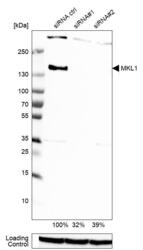
- Western blot analysis in U-251MG cells transfected with control siRNA, target specific siRNA probe #1 and #2, using Anti-MKL1 antibody. Remaining relative intensity is presented. Loading control: Anti-GAPDH.
- Sample type
- Human
- Protocol
- Protocol

Supportive validation
- Submitted by
- Atlas Antibodies (provider)
- Main image
- Experimental details
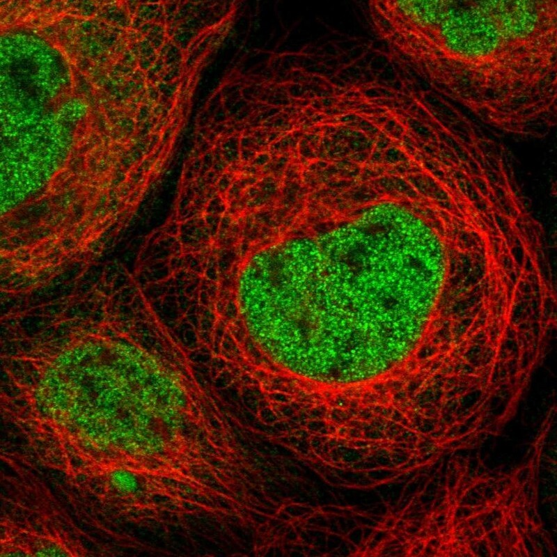
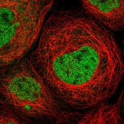
- Immunofluorescent staining of human cell line A-431 shows localization to nucleoplasm.
- Sample type
- Human