 Explore
 Validate
 Learn
Learn Western blot
 Flow cytometry
Antibody data
- Antibody Data
- Antigen structure
- References [3]
- Comments [0]
- Validations
- Western blot [1]
- Immunohistochemistry [2]
- Other assay [1]
Submit
Validation data
Reference
Comment
Report error
- Product number
- MA3-903 - Provider product page

- Provider
- Invitrogen Antibodies
- Product name
- HCN4 Monoclonal Antibody (SHG 1E5)
- Antibody type
- Monoclonal
- Antigen
- Synthetic peptide
- Description
- MA3-903 detects HCN4 from human, mouse, and rat samples. MA3-903 has been successfully used in Western blotting and immunohistochemistry procedures. By Western blot MA3-903 detects a ~132 KDa band representing HCN4. The MA3-903 immunogen is a synthetic peptide sequence corresponding to residues S H G S L L L P P A S S P P P P Q V P Q R R G T P P L T P G R L T Q D L K L of HCN4. MA3-903 clone SHG 1E5 is a rat monoclonal hybridoma. This antibody was produced by immunizing a rat, isolating the spleen cells, creating the hybridoma and successively injecting these cells into a mouse to produce ascites.
- Reactivity
- Human, Mouse, Rat
- Host
- Rat
- Isotype
- IgG
- Antibody clone number
- SHG 1E5
- Vial size
- 100 μL
- Concentration
- Conc. Not Determined
- Storage
- -20°C, Avoid Freeze/Thaw Cycles
Submitted references Thalamocortical neurons display suppressed burst-firing due to an enhanced Ih current in a genetic model of absence epilepsy.
HCN channels are expressed differentially in retinal bipolar cells and concentrated at synaptic terminals.
Hyperpolarization-activated channels HCN1 and HCN4 mediate responses to sour stimuli.
Cain SM, Tyson JR, Jones KL, Snutch TP
Pflugers Archiv : European journal of physiology 2015 Jun;467(6):1367-82
Pflugers Archiv : European journal of physiology 2015 Jun;467(6):1367-82
HCN channels are expressed differentially in retinal bipolar cells and concentrated at synaptic terminals.
Müller F, Scholten A, Ivanova E, Haverkamp S, Kremmer E, Kaupp UB
The European journal of neuroscience 2003 May;17(10):2084-96
The European journal of neuroscience 2003 May;17(10):2084-96
Hyperpolarization-activated channels HCN1 and HCN4 mediate responses to sour stimuli.
Stevens DR, Seifert R, Bufe B, Müller F, Kremmer E, Gauss R, Meyerhof W, Kaupp UB, Lindemann B
Nature 2001 Oct 11;413(6856):631-5
Nature 2001 Oct 11;413(6856):631-5
No comments: Submit comment
Supportive validation
- Submitted by
- Invitrogen Antibodies (provider)
- Main image

- Experimental details
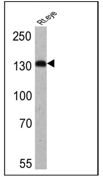
- Western blot analysis of HCN4 was performed by loading 25 µg of rat eye lysate onto an SDS polyacrylamide gel. Proteins were transferred to a PVDF membrane and blocked at 4ºC overnight. The membrane was probed with a HCN4 monoclonal antibody (Product # MA3-903) at a dilution of 1:200 overnight at 4°C, washed in TBST, and probed with an HRP-conjugated secondary antibody for 1 hr at room temperature in the dark. Chemiluminescent detection was performed using Pierce ECL Plus Western Blotting Substrate (Product # 32132). Results show a band at ~132 kDa.
Supportive validation
- Submitted by
- Invitrogen Antibodies (provider)
- Main image
- Experimental details
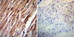
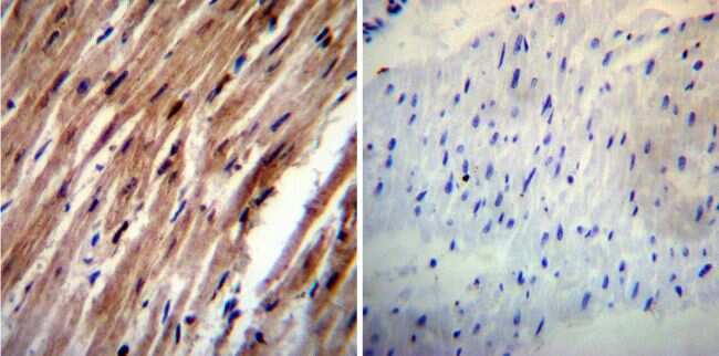
- Immunohistochemistry was performed on normal deparaffinized Human heart tissue tissues. To expose target proteins, heat induced antigen retrieval was performed using 10mM sodium citrate (pH6.0) buffer, microwaved for 8-15 minutes. Following antigen retrieval tissues were blocked in 3% BSA-PBS for 30 minutes at room temperature. Tissues were then probed at a dilution of 1:20 with a rat monoclonal antibody recognizing HCN4 (Product # MA3-903) or without primary antibody (negative control) overnight at 4°C in a humidified chamber. Tissues were washed extensively with PBST and endogenous peroxidase activity was quenched with a peroxidase suppressor. Detection was performed using a biotin-conjugated secondary antibody and SA-HRP, followed by colorimetric detection using DAB. Tissues were counterstained with hematoxylin and prepped for mounting.
- Submitted by
- Invitrogen Antibodies (provider)
- Main image
- Experimental details
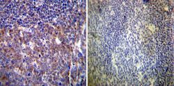
- Immunohistochemistry was performed on normal deparaffinized Human tonsil tissue tissues. To expose target proteins, heat induced antigen retrieval was performed using 10mM sodium citrate (pH6.0) buffer, microwaved for 8-15 minutes. Following antigen retrieval tissues were blocked in 3% BSA-PBS for 30 minutes at room temperature. Tissues were then probed at a dilution of 1:20 with a rat monoclonal antibody recognizing HCN4 (Product # MA3-903) or without primary antibody (negative control) overnight at 4°C in a humidified chamber. Tissues were washed extensively with PBST and endogenous peroxidase activity was quenched with a peroxidase suppressor. Detection was performed using a biotin-conjugated secondary antibody and SA-HRP, followed by colorimetric detection using DAB. Tissues were counterstained with hematoxylin and prepped for mounting.

Supportive validation
- Submitted by
- Invitrogen Antibodies (provider)
- Main image
- Experimental details
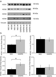
- Fig. 4 HCN protein isoforms are differentially expressed in GAERS VB thalamus. a Representative Western blots for HCN proteins from adult NEC and GAERS VB samples. Primary antibodies to detect each of the HCN channel isoforms were compared to vinculin in order to normalize the expression data. b Mean Western blot densitometry data for HCN1-4 normalized to vinculin for NEC ( n = 12 samples (six animals); black columns ) and GAERS ( n = 12 samples (six animals); grey columns ) VB samples
