Explore
 Validate
 Learn
Learn Western blot
 Immunocytochemistry
Antibody data
- Antibody Data
- Antigen structure
- References [1]
- Comments [0]
- Validations
- Immunocytochemistry [2]
- Immunohistochemistry [1]
- Flow cytometry [2]
- Other assay [3]
Submit
Validation data
Reference
Comment
Report error
- Product number
- 45-6300 - Provider product page

- Provider
- Invitrogen Antibodies
- Product name
- FXN Monoclonal Antibody (18A5DB1)
- Antibody type
- Monoclonal
- Antigen
- Other
- Description
- Positive controls: Recombinant Human Frataxin; normal Human fibroblasts (MRC5); HL60 cells. This antibody gave a positive result in IHC in the following FFPE tissue: Human colon adenocarcinoma.
- Reactivity
- Human
- Host
- Mouse
- Isotype
- IgG
- Antibody clone number
- 18A5DB1
- Vial size
- 100 μg
- Concentration
- 1 mg/mL
- Storage
- 4°C, do not freeze
Submitted references Long intronic GAA repeats causing Friedreich ataxia impede transcription elongation.
Punga T, Bühler M
EMBO molecular medicine 2010 Apr;2(4):120-9
EMBO molecular medicine 2010 Apr;2(4):120-9
No comments: Submit comment
Supportive validation
- Submitted by
- Invitrogen Antibodies (provider)
- Main image
- Experimental details
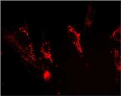
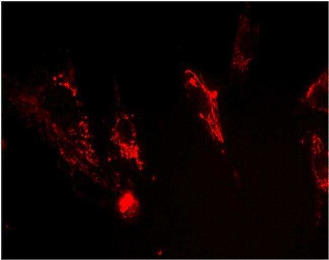
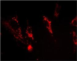
- Immunocytochemical analysis of FXN in MRC5 using a FXN Monoclonal antibody (Product # 45-6300) at a concentration of 0.5 µg/mL. The normal Human fibroblasts (MRC5) were fixed and permeabilized, and detected using an AlexaFluor® 594-conjugated-goat-anti-mouse IgG1 isotype specific secondary antibody (2 µg/mL).
- Submitted by
- Invitrogen Antibodies (provider)
- Main image
- Experimental details
- Immunocytochemical analysis of FXN in MRC5 using a FXN Monoclonal antibody (Product # 45-6300) at a concentration of 0.5 µg/mL. The normal Human fibroblasts (MRC5) were fixed and permeabilized, and detected using an AlexaFluor® 594-conjugated-goat-anti-mouse IgG1 isotype specific secondary antibody (2 µg/mL).

Supportive validation
- Submitted by
- Invitrogen Antibodies (provider)
- Main image
- Experimental details
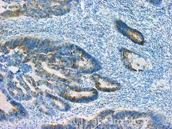
- Immunohistochemical analysis of FXN in Human colon adenocarcinoma tissue sections using a FXN Monoclonal Antibody (Product # 45-6300).

Supportive validation
- Submitted by
- Invitrogen Antibodies (provider)
- Main image

- Experimental details
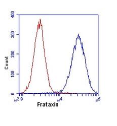
- Flow cytometric analysis of FXN in HL60 cells using an FXN monoclonal antibody (Product # 45-6300) at 1 µg/mL is depicted by the blue line. The red line indicates an isotype control antibody.

- Submitted by
- Invitrogen Antibodies (provider)
- Main image
- Experimental details
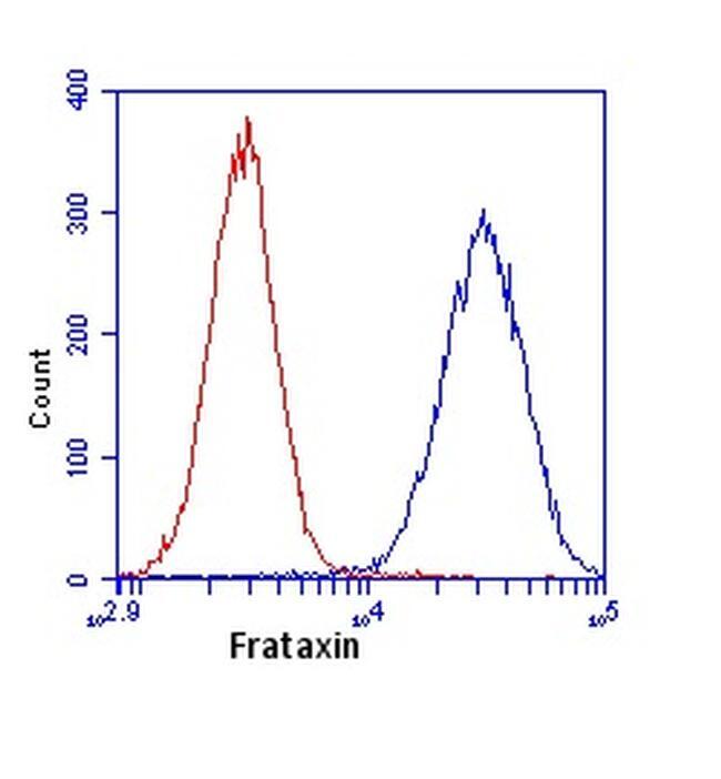
- Flow cytometric analysis of FXN in HL60 cells using an FXN monoclonal antibody (Product # 45-6300) at 1 µg/mL is depicted by the blue line. The red line indicates an isotype control antibody.
Supportive validation
- Submitted by
- Invitrogen Antibodies (provider)
- Main image
- Experimental details
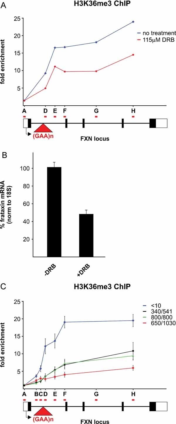
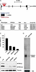
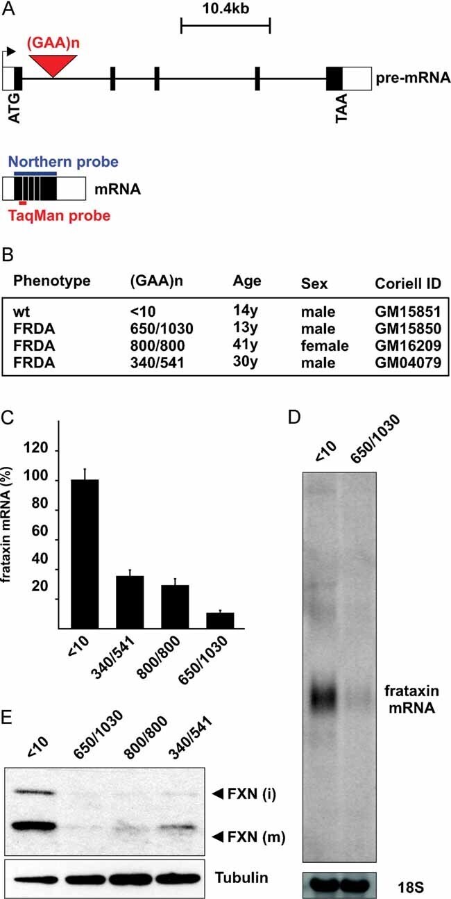
- Figure 1 Pathogenic GAA repeat expansions reduce FXN mRNA and protein levels Schematic view of the human FXN gene (ENSG00000165060). Exons and untranslated regions are depicted as black and white boxes, respectively. The red arrowhead indicates localization of the GAA repeats. Northern blot and TaqMan probes annealing to the FXN mRNA are indicated as blue and red lines, respectively. Cell lines used throughout this study. All cell lines were obtained from Coriell Cell Repositories. Reduced accumulation of FXN mRNA in FRDA cells as determined by qRT-PCR. Values for FXN mRNA were normalized to 18S rRNA. Northern blot showing that no major alternatively spliced FXN isoforms exist and that mRNA levels are reduced in FRDA cells. The Northern blot was probed with a human FXN cDNA probe. 18S served as a loading control. Western blot showing reduced accumulation of FXN protein in FRDA cells. Intermediate (i) and mature (m) forms of the FXN proteins are indicated.
- Submitted by
- Invitrogen Antibodies (provider)
- Main image
- Experimental details
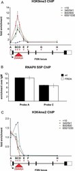
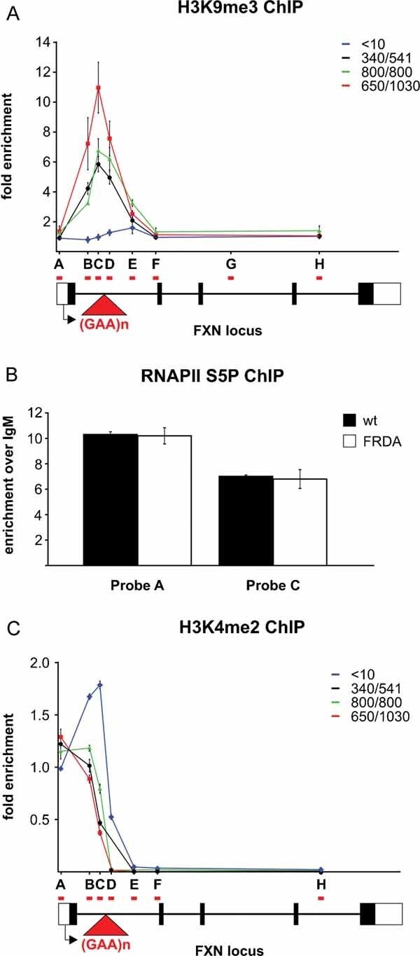
- Figure 3 H3K9 methylation is locally confined in FRDA cells and does not affect marks of transcription initiation ChIP assay to profile H3K9me3 along the FXN gene. The profile for wild type is shown in blue (
- Submitted by
- Invitrogen Antibodies (provider)
- Main image
- Experimental details
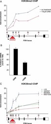
- Figure 4 Expanded GAA repeats affect H3K36 trimethylation Treatment of wild-type cells with the transcription elongation inhibitor DRB reduces the H3K36me3 signal along the FXN gene locus. Wild-type (GM15851) cells were treated with 115 uM DRB for 5 h before ChIP with anti-H3K36me3 antibody. Location of qPCR probes and fold enrichment is presented as in Fig 3A . Reduced accumulation of FXN mRNA after DRB treatment in wild-type (GM15851) cells. RNA was isolated from the same cells as used for the ChIP shown in (A). ChIP assay to profile H3K36me3 along the FXN gene in wild type and different FRDA cell lines. The profile for wild type is shown in blue (