 Explore
 Validate
 Learn
Learn Western blot
 Immunohistochemistry
Antibody data
- Antibody Data
- Antigen structure
- References [0]
- Comments [0]
- Validations
- Immunohistochemistry [1]
Submit
Validation data
Reference
Comment
Report error
- Product number
- AF5154 - Provider product page

- Provider
- R&D Systems
- Product name
- Human TAFA1/FAM19A1 Antibody
- Antibody type
- Polyclonal
- Description
- Immunogen affinity purified. Detects human TAFA1/FAM19A1 in direct ELISAs and Western blots. In direct ELISAs, approximately 15% cross-reactivity with recombinant human (rh) TAFA3 and rhTAFA4 is observed, less than 10% cross-reactivity with rhTAFA2 is observed and less than 1% cross-reactivity with rhTAFA5 is observed.
- Reactivity
- Human
- Host
- Goat
- Conjugate
- Unconjugated
- Antigen sequence
NP_998774- Isotype
- IgG
- Vial size
- 100 ug
- Concentration
- LYOPH
- Storage
- Use a manual defrost freezer and avoid repeated freeze-thaw cycles. 12 months from date of receipt, -20 to -70 °C as supplied. 1 month, 2 to 8 °C under sterile conditions after reconstitution. 6 months, -20 to -70 °C under sterile conditions after reconstitution.
No comments: Submit comment
Supportive validation
- Submitted by
- R&D Systems (provider)
- Main image
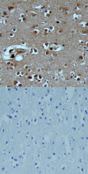
- Experimental details
- TAFA1/FAM19A1 in Human Brain. TAFA1/FAM19A1 was detected in immersion fixed paraffin-embedded sections of human brain (cortex) using Goat Anti-Human TAFA1/FAM19A1 Antigen Affinity-purified Polyclonal Antibody (Catalog # AF5154) at 15 µg/mL overnight at 4 °C. Tissue was stained using the Anti-Goat HRP-DAB Cell & Tissue Staining Kit (brown; Catalog # CTS008) and counterstained with hematoxylin (blue). Lower panel shows a lack of labeling when primary antibodies are omitted and tissue is stained only with secondary antibody followed by incubation with detection reagents. View our protocol for Chromogenic IHC Staining of Paraffin-embedded Tissue Sections.
