Explore
 Validate
 Learn
Learn Western blot
 Immunohistochemistry
Antibody data
- Antibody Data
- Antigen structure
- References [1]
- Comments [0]
- Validations
- Immunohistochemistry [1]
- Flow cytometry [2]
- Other assay [1]
Submit
Validation data
Reference
Comment
Report error
- Product number
- PA5-26069 - Provider product page

- Provider
- Invitrogen Antibodies
- Product name
- BPI Polyclonal Antibody
- Antibody type
- Polyclonal
- Antigen
- Synthetic peptide
- Reactivity
- Human
- Host
- Rabbit
- Isotype
- IgG
- Vial size
- 400 μL
- Concentration
- 2 mg/mL
- Storage
- Store at 4°C short term. For long term storage, store at -20°C, avoiding freeze/thaw cycles.
Submitted references The Mucosal Antibacterial Response Profile and Fecal Microbiota Composition Are Linked to the Disease Course in Patients with Newly Diagnosed Ulcerative Colitis.
Magnusson MK, Strid H, Isaksson S, Simrén M, Öhman L
Inflammatory bowel diseases 2017 Jun;23(6):956-966
Inflammatory bowel diseases 2017 Jun;23(6):956-966
No comments: Submit comment
Supportive validation
- Submitted by
- Invitrogen Antibodies (provider)
- Main image
- Experimental details
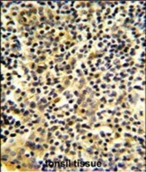
- Immunohistochemistry analysis of BPI in formalin-fixed and paraffin-embedded human tonsil tissue. Samples were incubated with BPI polyclonal antibody (Product # PA5-26069) which was peroxidase-conjugated to the secondary antibody, followed by DAB staining. This data demonstrates the use of this antibody for immunohistochemistry; clinical relevance has not been evaluated.

Supportive validation
- Submitted by
- Invitrogen Antibodies (provider)
- Main image
- Experimental details
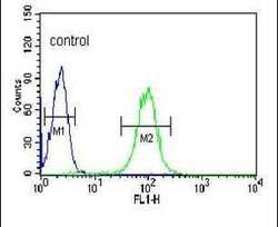
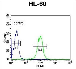
- Flow cytometry analysis of HL-60 cells using a BPI polyclonal antibody (Product # PA5-26069) (right) compared to a negative control cell (left) at a dilution of 1:10-50, followed by a FITC-conjugated goat anti-rabbit antibody
- Submitted by
- Invitrogen Antibodies (provider)
- Main image
- Experimental details
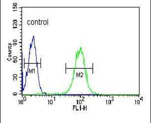
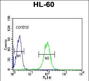
- Flow cytometry of BPI in HL-60 cells (right histogram). Samples were incubated with BPI polyclonal antibody (Product # PA5-26069) followed by FITC-conjugated goat-anti-rabbit secondary antibody. Negative control cell (left histogram).
Supportive validation
- Submitted by
- Invitrogen Antibodies (provider)
- Main image
- Experimental details
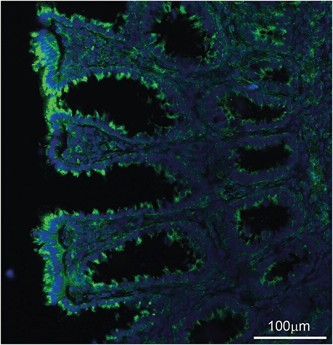
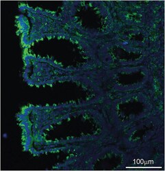
- FIGURE 4. Localization of BPI in the epithelial cell layer and crypts. Biopsy specimens from rectum stained with polyclonal rabbit-anti-BPI-IgG followed by goat-anti-rabbit-AlexaFluor488. Sections were mounted with 4', 6-Diamidino-2-Phenylindole Dihydrochloride (DAPI) (chromosome stain) and visualized at x20 magnification, green = BPI stain, blue = nuclear stain (DAPI). A representative staining from a patient with newly diagnosed UC is shown.