Explore
 Validate
 Learn
Learn Western blot
 Immunocytochemistry
Antibody data
- Antibody Data
- Antigen structure
- References [1]
- Comments [0]
- Validations
- Immunocytochemistry [1]
- Other assay [1]
Submit
Validation data
Reference
Comment
Report error
- Product number
- 702076 - Provider product page

- Provider
- Invitrogen Antibodies
- Product name
- SLC6A4 Recombinant Rabbit Monoclonal Antibody (16H2L7)
- Antibody type
- Monoclonal
- Antigen
- Synthetic peptide
- Reactivity
- Human, Mouse, Rat
- Host
- Rabbit
- Isotype
- IgG
- Antibody clone number
- 16H2L7
- Vial size
- 100 µg
- Concentration
- 0.5 mg/mL
- Storage
- Store at 4°C short term. For long term storage, store at -20°C, avoiding freeze/thaw cycles.
Submitted references The effect of citalopram treatment on amyloid-β precursor protein processing and oxidative stress in human hNSC-derived neurons.
Elsworthy RJ, Crowe JA, King MC, Dunleavy C, Fisher E, Ludlam A, Parri HR, Hill EJ, Aldred S
Translational psychiatry 2022 Jul 18;12(1):285
Translational psychiatry 2022 Jul 18;12(1):285
No comments: Submit comment
Supportive validation
- Submitted by
- Invitrogen Antibodies (provider)
- Main image

- Experimental details
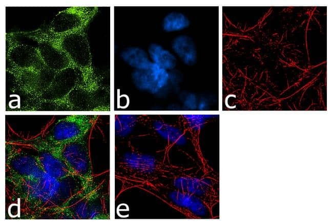
- For immunofluorescence analysis, IMR-32 cells were fixed and permeabilized for detection of endogenous SERT using Anti- SERT Recombinant Rabbit Monoclonal Antibody (Product # 702076, 2 µg/mL) and labeled with Goat anti-Rabbit IgG (H+L) Superclonal™ Secondary Antibody, Alexa Fluor® 488 conjugate (Product # A27034, 1:2000). Panel a) shows representative cells that were stained for detection and localization of SERT protein (green), Panel b) is stained for nuclei (blue) using SlowFade® Gold Antifade Mountant with DAPI (Product # S36938). Panel c) represents cytoskeletal F-actin staining using Alexa Fluor® 555 Rhodamine Phalloidin (Product # R415, 1:300). Panel d) is a composite image of Panels a, b and c clearly demonstrating membrane localization of SERT. Panel e) represents control cells with no primary antibody to assess background. The images were captured at 60X magnification.
Supportive validation
- Submitted by
- Invitrogen Antibodies (provider)
- Main image
- Experimental details
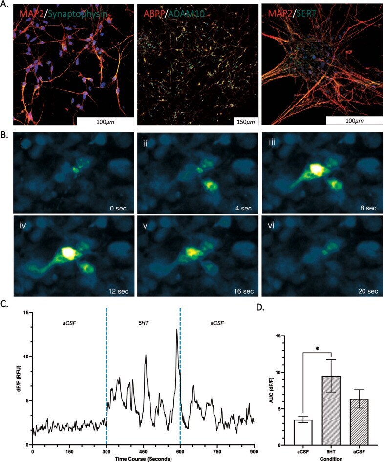
- Representative microscopy images of cultured cells with ICC staining (A) and calcium imaging (B & C) ( n = 3). A Mature neuronal staining positive for MAP2, Synaptophysin, SERT, ADAM10 and AbetaPP (AX0018, d45) with nucleus staining for DAPI (blue). B PSEN1 (L286V) fluoro-4-am calcium transient measured in neurons (day 42). C Signal intensity was used to calculate neuronal activity (DF/F) under basal (aCSF), during stimulation with 5HT and a wash off period (aCSF) in 5-min intervals ( n = 3). D Area under the curve plotted for each condition to analyse neuronal activity (3ROIs, n = 3). Data normality (p > 0.05) and S.D. Variation (p < 0.05) confirmed assumptions for ANOVA analysis with multiple comparison corrections applied. *Significantly different to treatment condition ( p < 0.05).